| |

What is the full form of CT-SCANCT-SCAN: Computed Tomography Scan
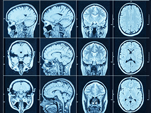
CT-SCAN stands for Computed Tomography Scan. A computed tomography scan, often known as a CAT or computerized tomography scan, is a diagnostic imaging method requiring inside images of the body. The individuals who perform CT scans are known as radiologists or radiography technologists. CT scanners use a spinning X-ray pipe and a stack of sensors mounted in a carriage to measure the help to aid of X-rays by various tissues within the person. Tomographic reconstruction algorithms are then used to analyze the many X-ray data gathered from multiple angles on a computer to create tomographic (cross-sectional) images (virtual "slices") of a body. Clients who need a CT scan but cannot endure an MRI do so because they have metallic devices or cardioverters. HistoryThe history of computed tomography is the primary topic. The Radon transforms mathematical theory that can be traced back to at least 1917 in the development of X-ray computed tomography. A U.S. patent for a "radiant energy system for probing specified parts of inner objects hidden by dense material" was granted to William H. Oldendorf in October 1963. Godfrey 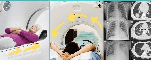
Hounsfield created the first CT scanner we could use for commercial purposes in 1972. Medical UseThe introduction of CT scanning in the mid-1960s shows it to be a robust optical technique. While the most common application of CT is for medical diagnosis, we can also use it to create images of inanimate objects. As a supplement to traditional X-ray imaging and medical ultrasonography, CT has grown in importance since its inception in the 1970s. It has become increasingly common for disease control and monitoring, including CT colonography for populations at high risk of colorectal cancer and single heart imaging for individuals at risk of heart disease. 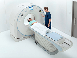
Many institutions provide full-body scans to the general public. However, this practice defies the recommendations and official stance of many professional organizations in the industry, primarily because of the radiation dose used. Over the past 20 years, the utilization of CT scans has substantially expanded in many nations. In the United States, we did an estimated 72 million scans in 2007 and more than 80 million in 2015. Industrial UseIndustrial CT scanning, also known as industrial computed tomography, is a technique that makes 3D models of both the inside and outside components using X-ray technology. Some significant applications for CT scanning include defect identification, fault diagnosis, metrology, assembling analyzation, image-based finite difference methods, and information extraction. The imaging and preservation of museum objects also use CT scanning. CT scanning has been used during transport security (central security at the airport) using object tracking visual recognition methodologies. 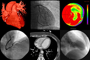
It is presently included in a substances session for explosive materials identification CTX (an act of terrorism device) and has also been regarded for computer-controlled transportation security scanning. Such techniques are meant to identify specific attack goods based on the 3D presence (e.g. guns, knives, liquid containers). Heathrow Airport aims for an initial roll on December 1, 2022, and the TSA expended $781.2 million over an agreement for further than 1,000 readers, prepared to go active in the summertime, to implement its use in airport security, which they launched at Shannon Airport in March 2022. Use of GeologyX-ray Geoscience investigations employ CT to expose the contents of drill cores swiftly. In Computed tomography, solid materials like pyrite and barite seem more vital, although a few lesser solid substances like clay look dimmer. Utilization of Cultural HeritageMicro-CT and X-ray CT can be employed to protect and sustain cultural history artefacts. Direct investigation and inspection can harm many delicate objects and, over time, destroy them. Restorers and scientists can securely use CT scans to ascertain the material makeup of the things they are investigating, such as ink distribution along a scroll's layers. Such scanners have proved ideal for studies focusing on the operation of the Antikythera mechanism or the text concealed within the En-Gedi Scroll's burnt outer layers. 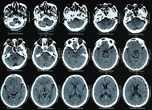
They are not suitable for all objects involved in these study concerns, as some relics, such as the Herculaneum papyri, show slight internal variation in the material makeup. After these artefacts have been scanned, we can use optimization algorithms to look within them. Researchers did it with the digital unravelling of the En-Gedi scroll and the papyri from Herculaneum. More modern relics, such as still-sealed medieval communications that used the letter locking technique (complicated folding and cutting that formed a "tamper-evident locking system), have been proven to be amenable to analysis using micro-CT. Imaging the contents of sarcophagi or ceramics are more examples of use cases in archaeology. AdvantagesCompared to conventional two-dimensional medical radiography, CT scanning has several benefits.
Adverse EffectsCancerThe radiation in CT scans can harm DNA molecules in body cells, resulting in cancer from radiation exposure. Variable radiation dosages are received during CT scans. CT scans can have a dose 100-1,000 times higher than traditional X-rays compared to the lowest dose x-ray procedures, and a head CT has a lower amount than a lumbar spine x-ray. By contrasting the lowest-dose x-ray techniques (chest x-ray) with the highest-dose CT procedures, articles in the media frequently overestimate the relative dose of CT. Typically, a standard abdominal CT exposes the patient to a radiation dose equivalent to three years of background radiation on average. 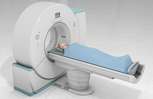
The large cumulative doses of more than 100 mSv to individuals getting recurring CT scans within a short period of 1 to 5 years have come to light in recent research on 2.5 million patients and 3.2 million patients. There is alarmingly little evidence that the current high incidence of scans is related to better health outcomes, according to several experts, who also add that CT scans are known to be "overused." However, a recent article that examined the data of patients who received high cumulative dosages of the drug revealed a high level of appropriate use. It raises a crucial problem regarding the patients' likelihood of developing cancer. Additionally, a previously unknown and significant discovery is that some patients received more than 100 mSv of radiation from a single CT scan. Initial assessments of CT danger are based partly on radiation doses comparable to those of nuclear industry workers and individuals residing during the blasts of the atomic bombs in Japan following World War II. According to some specialists, approximately three to five per cent of all cancers are caused by medical imaging in future. According to an Australian study of 10.9 million persons, radiation exposure was mainly to blame for the higher cancer incidence in this cohort following CT scan exposure. In this group, scientists detected excess cancer after 1,800 CT scans. The absolute risk increases to 40.05 per cent following a CT if the lifetime risk of acquiring cancer is 40 per cent. According to several studies, reports claiming an increased risk of cancer from standard body CT scan dosages suffer from significant methodological flaws and several implausible findings. And they are leading to the conclusion that there is no proof that such modest doses have any adverse long-term effects. According to one study, using CT scans may have contributed to as many as 0.4 per cent of cancer cases in the United States in 2007, and this number may have gone as high as 1.5 to 2 per cent. There is disagreement about whether the modest doses of radiation used in CT scans harm. Thus, some people dispute this estimate. Lower radiation doses are used in several situations, such as investigating renal colic. Age significantly affects a person's future chance of developing cancer. A one-year-abdomen old's CT is estimated to carry a lifetime cancer mortality risk of 0.1 per cent, or one scan every 1000. With significantly lower danger in the elderly, the risk for someone 40 years old is half that of someone 20. According to the International Commission on Radiological Protection, the chance of developing cancer before the age of 20 increases from 0.03 per cent to 0.04 per cent when a foetus is exposed to 10 mGy (a unit of radiation exposure) (for reference, a CT pulmonary angiogram reveals a foetus to 4 mGy). A 2012 analysis found no link between medical radiation and children's cancer risk despite recognizing a relationship. As of 2007, most CT scan manufacturers had this function built in, allowing different settings for CT scans to be performed for lower exposure in children. Additionally, certain illnesses may mandate that kids undergo several CT scans. According to recent research, we should warn parents about the dangers of children's CT scans. Radiation EffectsRadiobiology The majority of harmful health effects from radiation exposure can be divided into two categories:
A single abdominal CT of 8 mSv is thought to increase lifetime cancer risk by 0.05 per cent, or 1 in 2,000. The radiation dosage of a CT scan is a crucial factor in the decision of medical imaging in pregnancy because foetuses are more susceptible to radiation exposure. Campaigns In Society And CultureThe Society for Pediatric Radiology established the Alliance for Radiation Safety in Pediatric Imaging in response to growing public concern and the development of best practices. The Society for Pediatric Radiology created and launched the Image Gently Campaign, which aims to maintain high-quality imaging studies while using the lowest doses and best radiation safety practices on paediatric patients. Collaborating with the American Society of Radiologic Technologists, the American College of Radiology, and the American Association of Physicists in Medicine. 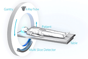
A rising number of international professional medical organizations have supported and applied for this project, which has also garnered help and support from businesses that provide equipment used in radiology. The American Society of Radiologic Technologists has launched a similar campaign called Image Wisely to address this issue in the adult population in response to the success of the Image Gently campaign. The World Health Organization and the United Nations' International Atomic Energy Agency (IAEA) have worked in this field. They have ongoing initiatives to extend best practices and reduce patient radiation doses. ConclusionNearly half of the overall per-capita dose rate from radiologic and nuclear medicine procedures was accounted for by the projected 72 million scans that were carried out in the United States in 2007, according to estimates. Six to eleven per cent of CT scans are performed on children, up seven to eight times since 1980. Asia and Europe have each experienced similar growth. In Calgary, Canada, 12.1% of patients who arrive at the emergency department with an urgent complaint had a CT scan, most frequently of the head or abdomen. However, the percentage of patients who had CT varied significantly depending on ER doctor who treated them, ranging from 1.8 per cent to 25 per cent.
Next TopicFull Forms List
|
 For Videos Join Our Youtube Channel: Join Now
Feedback
- Send your Feedback to [email protected]
Help Others, Please Share


