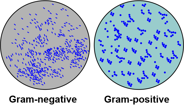
Difference Between Gram-Positive Bacteria and Gram-Negative BacteriaThere are two types of bacterial infections caused by two different types of bacteria - gram-positive and gram-negative. The two types of bacteria are differentiated based on a microbial technique called gram staining. The technique involves staining the bacterial cell with certain dyes. Both types of bacteria have different cell wall structures, and the ability of the bacterial cell wall to retain the dye color differentiates gram-positive from gram-negative bacteria. 
Gram Staining Technique -The technique is relatively quick and helps health practitioners identify the bacteria at the site of infection. According to this technique, the two types of bacteria are stained, red or purple, based on their type of cell wall. The technique was developed by a famous bacteriologist, Hans Christian Gram, in 1882. The technique was named after his name, and he developed this technique while working on pneumonia. Bacterial cultures are used to diagnose the type of bacteria present. The bacterial culture is put on a glass slide and observed under a microscope. Upon the identification of the color of the bacterial culture, they are differentiated as gram-positive or gram-negative. This technique is quicker than most other bacterial culture techniques. However, it is inefficient to diagnose the specific bacterium present in the culture, which is the main cause of the disease. It is used as a first step in giving the right direction for identifying bacteria. Many infections, like pneumonia, urinary tract infections, etc., are diagnosed using gram staining. 
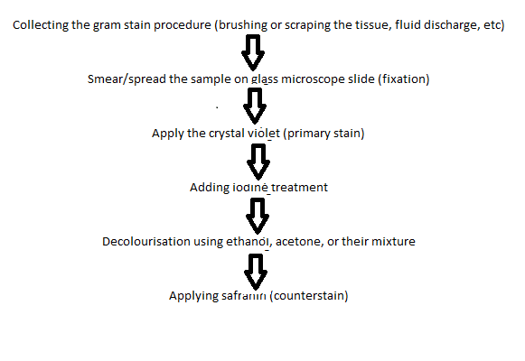
The sample of bacteria is taken from the site of infection. The sample can be in the form of a swab or mucus. It is then contained in a sterile container to send to the laboratory. The medical laboratory scientist put the samples on the glass slide and applied a series of stains to the bacterial culture. After performing all the technique steps, the cells are observed under the microscope. The different steps in the gram staining technique are as follows -
Gram staining procedure -
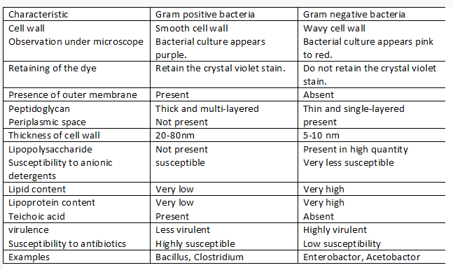
Observation and results -After performing the staining procedure, the scientists observe the culture's color under the microscope. If the bacterial culture appears purple, then it is gram-positive bacteria. If the bacterial culture appears pink to red, it is gram-negative bacteria. The results are due to the bacteria's cell wall structure difference. The difference between gram-positive and gram-negative bacteria
Conclusion -The major difference between gram-positive and gram-negative bacteria is the difference in their cell walls. The gram stain dye is retained in the cell wall due to the thickness of peptidoglycan present in gram-positive bacteria. Gram-negative bacteria lack peptidoglycan; therefore, they do not retain the gran stain and appear red to pink after decolorization with ethanol. The major dye responsible for staining the bacteria is crystal violet stain. Crystal violet is purple and gives the characteristic color to the bacterial culture, which helps differentiate the two types of gram bacteria.
Next TopicDifference between
|
 For Videos Join Our Youtube Channel: Join Now
Feedback
- Send your Feedback to [email protected]
Help Others, Please Share


