| |

What is the full form of ECGECG: ElectrocardiogramECG stands for Electrocardiogram. It refers to a test that is performed to check the electrical activity of the heart. Normal electrical activity indicates that the heart is working normally. 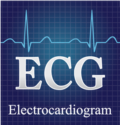
ECG records the electrical impulses when the heart is beating. These impulses are recorded on a moving strip of paper or on a screen, i.e., it shows the heart's electrical activity as line tracings on paper. The spikes and dips that appear in the tracings are called waves. So, a normal ECG line tracing consists of waves which are as follows: P Wave: It is the first upward movement of the ECG tracing. It shows the electrical activity of the upper heart chambers. QRS Complex: It consists of three waves that begin with a downward deflection (Q), then a large upward deflection (R), and then a downwards S wave. It shows the electrical activity of the lower heart chambers. ST Segment: It appears as a straight line when the ventricle contracts without an electrical impulse. T Wave: It is a modest upwards wave that represents ventricular repolarization, i.e. when the lower heart chambers are preparing for the next muscle contraction. If is there any problem with hearts? Rhythm, there will be variations in the pattern of impulses or the graph. It is a painless procedure. It is performed by placing electrodes at specific places on your body, like arms, legs or chest. Why is an ECG needed?ECG can detect various heart-related problems. Some are listed below;
Types of ECG: There are three main types of ECG which are as follows; Resting ECG: It is carried out when the patient is lying down in a comfortable position. Stress or Exercise ECG: It is carried out when you are using a treadmill or exercise bike. Ambulatory ECG: The patient is required to wear a small machine at his or her waist. The patient may go home and resume his or her routine work. It helps monitor the heart for one or more days. RisksThe ECG technique is risk-free. As the test electrodes don't generate any electricity, electrical shock is not a worry. Just the heart's electrical activity is captured by the electrodes. You can feel a little discomfort as the electrodes are removed, similar to removing a bandage. Some individuals may get a mild rash where the patches are applied. How you prepareYou don't need to make any additional preparations to do a standard ECG. Tell your doctor about all prescription medications and dietary supplements you take. They frequently have an impact on an ECG's outcomes. What to anticipateAn ECG can be performed at a hospital or a doctor's office. BeforeYou could be required to put on a hospital gown. The doctor may remove any hair from the areas of your body where the electrodes will be implanted in order for the patches to stick. After you're prepared, you'll normally be asked to lie down on a bed or examination table. DuringUp to 12 sensors (electrodes) may be affixed to the chest and limbs during an ECG. The electrodes are wire-connected adhesive patches that cling to the skin. The electrical impulses that the heart utilizes to beat may be recorded. The data is first stored in a computer before being shown as waves on a screen or on paper. Throughout the exam, you can breathe, but you must remain still. Make sure you are prepared to lie motionless and are warm. Moving, speaking, or shivering might affect the test's outcomes. A typical ECG requires a few minutes. AfterAfter your ECG, you can usually resume your regular activities. ResultsYour doctor may go through the findings with you the day after your ECG or at your subsequent visit. A healthcare professional may get information from an ECG concerning the following: A heartbeat: Typically, the pulse may be checked to determine the heart rate. If your pulse is hard to feel, too rapid, or too irregular to count precisely, an ECG may be beneficial. An abnormally high heart rate (tachycardia) or an abnormally sluggish heart rate can both be found using an ECG (bradycardia). Heart rhythm: An ECG can identify unsteady heartbeats (arrhythmias). When any component of the heart's electrical system malfunctions, an arrhythmia may happen. Chest pains: An ECG might reveal signs of a present heart attack or one that has already occurred. The patterns on the ECG may aid in identifying the amount of damage to the heart as well as the specific area that has been harmed. Blood and oxygen are supplied to the heart: By doing an ECG while you are experiencing symptoms, your healthcare provider can determine whether your chest pain is being caused by reduced blood flow to the heart muscle. The shape of the heart varies: An ECG can tell you about heart problems, including enlarged hearts and other cardiac irregularities. You might require a second ECG or another test, such as echocardiography, if the results indicate a cardiac rhythm issue. Therapy is based on the root of your symptoms and indicators.
Next TopicFull Form
|
 For Videos Join Our Youtube Channel: Join Now
Feedback
- Send your Feedback to [email protected]
Help Others, Please Share


