| |

What is the Full Form of ERCPERCP: Endoscopic retrograde CholangiopancreatographyERCP stands for Endoscopic retrograde cholangiopancreatography. It is a method for diagnosing and treating specific issues with the biliary and pancreatic ductal systems that include the use of endoscopy & fluoroscopy. Most gastroenterologists with advanced training and ability conduct it. The doctor can use an endoscope to see into the duodenum and biliary tree and inject a contrast agent into the ducts of the pancreas and biliary tree to make them visible on radiography. 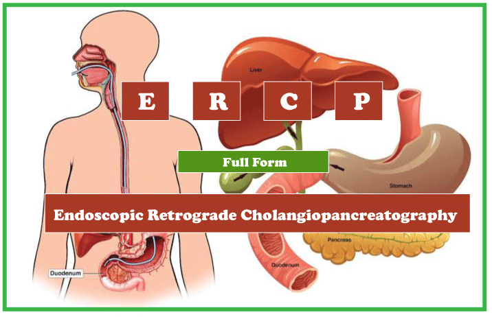
Gallstones, inflamed strictures (scars), leaks (after trauma and surgery), and cancer are among the disorders of the bile ducts and primary pancreatic ducts that are typically diagnosed and treated with ERCP. Although ERCP can be used for diagnostic and therapeutic purposes, it is currently seldom used without a therapeutic purpose due to the development of safer and more minimally invasive procedures like magnetic resonance cholangiopancreatography (MRCP) and endoscopic ultrasonography. Medical UsesDiagnosticThe following are indications for using the ERCP, especially if and when less invasive alternatives are insufficient or unsatisfactory:
Due to the increasing accessibility of safer diagnostic techniques, including endoscopic ultrasonography, CT, and MRI/MRCP, chronic pancreatitis is a contentious issue. Except when they result in bile duct blockage and jaundice, pancreatic tumours no longer constitute a reliable diagnostic indication for ERCP. A more secure and reliable diagnostic option is endoscopic ultrasonography. TherapeuticIn addition to the above diagnostic conditions, ERCP may be recommended if any of the following are necessary:
Contraindications
A background of iodinated contrast dye allergy or hypersensitivity to iodine-based contrast medium is not a contraindication to ERCP. Still, it should be addressed with your health practitioner. You should also inform your doctor if you are sensitive to iodine. As an alternate, contrast iodine-free substance ("dye") is then inserted gently into the duct system (pancreatic or biliary) before x-rays are obtained. ProcedureThe patient must be either asleep or put under anaesthesia. After that, an endoscope-a flexible camera-is entered via the mouth, moving through the oesophagus, stomach, pylorus, and duodenum, where the ampulla of Vater-the junction of the central bile duct and pancreatic duct-is located. The muscle valve known as the sphincter of Oddi regulates based on how wide the ampulla opens. With the endoscopic camera, the area may be seen immediately while various treatments are being carried out. A plastic catheter or cannula is introduced through the ampulla, and radiocontrast is administered into the pancreatic and bile ducts. To detect blockages or other lesions like stones, fluoroscopy is employed. When necessary, the sphincters of the bile ducts and ampulla can be opened (via the sphincterotomy) with an electric wire known as a sphincterotome to provide access so that gallstones can be removed, or other treatments can be administered. The removal of gallstones from the common bile duct using a basket or balloon and the placement of a plastic stent to help with bile drainage are two more ERCP procedures. Additionally, stents may be implanted after cannulating the pancreatic duct. In situations of pancreatitis, it is necessary to see the pancreatic duct. Although it has minimal benefit in evaluating pancreatitis or its consequences, ultrasound is typically the first test done after admission. The most used imaging method is called MD-CECT, or contrast-enhanced computed tomography. But magnetic resonance imaging (MRI) provides diagnostic skills comparable to computed tomography (CT), with additional inherent benefits such as the absence of ionizing radiation and excellent soft tissue characterization. Other specialized or auxiliary endoscopes may be utilized for ERCP in certain circumstances. In addition to balloon enteroscopy, the procedure may also involve mother-baby or SpyGlass cholangiocytes (to aid in diagnosis by directly observing the duct instead of simply acquiring X-ray pictures). RisksPost-ERCP pancreatitis (PEP) is one of the most common and dreaded side effects following endoscopic retrograde cholangiopancreatography (ERCP). The occurrence of PEP has indeed been calculated to range between 3.5 and 5% in earlier investigations. "Clinical pancreatitis involving amylase at least three times the maximum limit of normal at further than 24 hours following the surgery, requiring hospitalization or prolonging of planned stay" is what Cotton et al. classify as PEP. The length of the hospital stay is the primary factor in grading the severity of PEP. Technical aspects of the ERCP operation and patient-specific aspects are risk factors for PEP. The patient-related characteristics include female gender, younger age, and Sphincter of Oddi malfunction; the technical aspects include manipulation and injection of contrasts into the pancreatic duct, cannulation efforts lasting longer than five minutes, with biliary balloon sphincter dilatation. A previous history of PEP or pancreatitis considerably increases the danger for PEP to 17.8% and 5.5%, respectively, according to a comprehensive evaluation of clinical studies. Any gastroenterologist endoscopic surgery risks intestinal perforation, and performing a sphincterotomy increases that risk. Due to the anatomical placement of the second half of the duodenum, which lies behind the peritoneal structures of the abdomen, sphincterotomies-related perforations are retroperitoneal. Sphincterotomy carries a danger of bleeding as well. Through damage to friable hilar tumours or a guidewire piercing the bile duct wall and forming a biliary fistula, ERCP may cause hemobilia. The infrequent but potentially fatal sphincterotomy complication of delayed bleeding is made worse because many patients are sent home shortly after ERCP. Even though the anaphylactoid responses take place when you are in the hospital, there is a danger linked with the contrast dye for people who are allergic to substances containing iodine. Oversedation can cause vomiting, nausea, respiratory depression, hazardously low blood pressure, and other symptoms. Heart and lung issues, as well as the potentially fatal cholangitis infection of the bile duct, are other possible consequences (less than 1% of cases). The use of antibiotics before surgery has been shown to reduce the risk of cholangitis and septicemia. Although rare, ERCP complications can also be deadly. According to the Food and Drug Administration, hospital-acquired (i.e., nosocomial) infections involving carbapenem-resistant Enterobacteriaceae have been related to inadequately disinfected duodenoscopes since at least 2009. In 2013, the Virginia Mason Hospital in Seattle and the UCLA Health System in Los Angeles, Chicago, and Pittsburgh; all reported outbreaks. In February 2015, the FDA released a safety alert titled "Configuration of ERCP Duodenoscopes Might Impede Effective Cleaning", which was later amended in December 2015 and, most recently, in 2022 and advised disposable components. A relative bile deficit can cause fat malabsorption and inadequacies of fat-soluble vitamins. This is because bile is necessary for the digestive system's absorption and degradation of fat.
Next TopicFull Form
|
 For Videos Join Our Youtube Channel: Join Now
Feedback
- Send your Feedback to [email protected]
Help Others, Please Share


