| |

What is the Full Form of MRI in MedicalMRI: Magnetic Resonance ImagingMRI stands for Magnetic Resonance Imaging. It is a non-invasive image processing technique that generates three-dimensional images. It is often used in the identification, diagnosis, and monitoring of illnesses and their treatments. MRI is a modern technology that excites and detects changes in the axis of rotation of ions found in water that are present in living tissues of the human body. 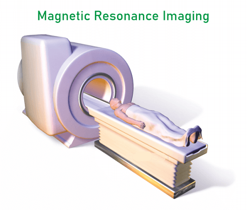
How does it work?Powerful magnets are used in MRI to create a strong magnetic field that compels body ions to align with it. The patient is pulsed with a radiofrequency current, which makes the electrons get excited and spin out of balance. This leads them to struggle against the magnetic field's lift. The energy released by charged particles when they align with the magnetic flux can be monitored by MRI detectors even when the electromagnetic field is shut off. The surroundings and the chemical interactions between the molecules affect how much energy is produced and how long it takes for the charged particles to align with the magnetic field. Depending on these magnetic properties, doctors can distinguish between various types of tissues. A patient is positioned inside a superconducting magnet to achieve an MRI image and should remain very still throughout the imaging process to prevent blurring the image. Some agents usually containing the element Gadolinium may be injected intravenously i.e. in the veins of a patient just before or during an MRI to speed up the rate at which charged particles realign with the magnetic field as a result image brightens as the ions readjust faster. Where is it used?MRI scanners are particularly appropriate for imaging the body's internal organs or surrounding tissues. They are clearly different from ultrasound imaging because they do not employ the harmful ionizing radiation of X-rays. MRI can scan the brain, spinal cord, and nerves significantly more clearly than standard X-rays and CT scans, as well as muscles, tendons, and ligaments. MRI is extensively used to examine knee and shoulder injuries. 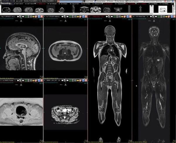
MRI can distinguish between white and gray matter within the brain and can also be used to diagnose haemorrhages and tumours. MRI does not use X-rays or other radiation, therefore is the widely imaging technique when imaging is required for diagnostic tests or therapy, especially for the brain. MRI, in contrast, is more costly than X-ray or Computerized tomography (CT Scan). MRI is used to examine brain structures and decide which areas of the brain are active and take in more oxygen during cognitive tasks. It is used to provide more understanding of multiple brain regions and may also provide a standard for evaluating the neurological status and neurosurgical risk. Risks associated with MRIThings to be kept in mind before doing MRI Scan:
Next TopicFull Form
|
 For Videos Join Our Youtube Channel: Join Now
Feedback
- Send your Feedback to [email protected]
Help Others, Please Share


