| |

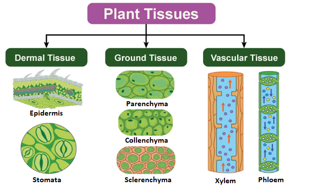
Difference Between Meristematic and Permanent TissuesIntroductionThe biological organizational level between cells and a whole organ is called a tissue in biology. Hence, tissue is often considered an amalgamation of comparable cells and their extracellular matrix from the same origin that work together to perform a certain function. Afterward, various tissues are functionally grouped to create organs. The French word "tissu," the past tense of the verb "to weave," is where the English term "tissue" originates. Histology, or histopathology when applied to illness, is the study of tissues. The "Father of Histology" is Xavier Bichat. Both plant anatomy and physiology study plant histology. The traditional methods for analyzing tissues are the paraffin block, in which the tissue is fixed and subsequently cut into sections, the histology dye, and the optical microscope. The level of detail that can be seen in tissues has increased as a result of advancements in immunofluorescence, electron microscopy, and the use of frozen tissue slices. These instruments make it possible to compare the traditional appearances of tissues in health and sickness, greatly improving medical diagnosis and prognosis. Plant TissuesThe epidermis, ground tissue, and vascular tissue are the three major systems used to classify tissues in plant anatomy.
Plant tissues may be further classified into two groups:
Meristematic TissuesMeristematic tissue, made up of actively dividing cells, causes the plant to grow longer and thicker. Only select plant areas, such as the tips of stems or roots, experience primary growth. Meristematic tissue may be seen in these areas. This kind of tissue's cells has thin cell walls and a general spherical to polyhedral to rectangular form. At first, the new cells generated by meristem are those of the meristem itself. Still, as they develop and increase, their properties gradually alter, and they differentiate into meristematic tissue components, being categorized as:
The main cell wall of meristematic tissue cells is formed of cellulose and is both thin and elastic. They don't have any intercellular gaps between them and are grouped closely together. Each cell has a large cell nucleus and a thick layer of cytoplasm. There are hardly any vacuoles in the dense protoplasm of meristematic cells. The meristematic cells are often rectangular, polygonal, or oval in form. Meristematic tissue cells have little need for intercellular gaps and a large nucleus since their role is to proliferate and increase the girth and length of the plant, not to store anything. They also contain minimal or no vacuoles. 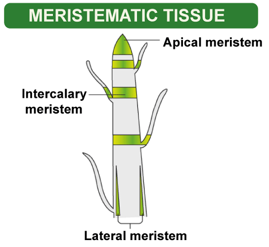
Permanent TissuesA set of meristematic tissue-derived live or dead cells that have lost the capacity to divide and have been fixedly positioned in the body of the plant are referred to as permanent tissues. Meristematic tissues that adopt a certain function lose their capacity to increase. Cellular differentiation is the process through which a permanent form, size, and function take hold. Several kinds of permanent tissues are created by differentiating meristematic tissue cells. Permanent tissues come in two varieties:
Simple Permanent TissueA collection of cells with the same origin, structure, and function makes up simple permanent tissue. They come in three categories:
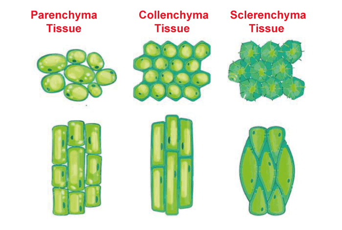
i) Parenchyma The majority of a material is referred to as parenchyma (Greek: para- "beside"; enchyma- "infusion" - "tissue"). It is made up of generally unspecialized living cells in plants that are often loosely packed and have thin cell walls, resulting in intercellular gaps between the cells of this tissue. They often have an isodiametric form. They have limited vacuoles, and sometimes they may even have none. Even if they do, the vacuole is still far smaller than in typical animal cells. In addition to supporting plants, this tissue also stores food. A unique kind of parenchyma called chlorenchyma accomplishes photosynthesis and includes chlorophyll. Aquatic plants' aerenchyma tissues, which contain substantial air spaces, provide them with the buoyancy they need to float on the water. Metabolic waste is present in Idioblasts, which are parenchyma cells. Prosenchyma, or fiber in the form of a spindle, is also found within these cells to sustain them, along with succulent parenchyma. Parenchyma tissues of xerophytes retain water. ii) Collenchyma Collenchyma, which means "gum infusion" in Greek, is a living tissue of the main body, similar to the parenchyma. While cells have thin walls, the corners where many cells converge include thickenings of cellulose, water, and pectin compounds (pectocellulosic). This tissue offers the plant tensile strength, and the cells are closely packed together with little intercellular gaps. The hypodermis of stems and leaves is where it mostly appears. Monocots and roots don't have it. On the stems of immature plants, collenchymatous tissue serves as a supporting tissue. It gives the plant body mechanical support, elasticity, and tensile strength. It facilitates the production of sugar and its storage as starch. It can withstand the shredding force of the wind and is found on the border of leaves. iii) Sclerenchyma Thick-walled, dead cells make up sclerenchyma, which is derived from the Greek words for "infusion" and "hard" (sclerous). Protoplasm is minimal. Due to the uniform distribution and high secretion of lignin, these cells have rigid and very thick secondary walls, which serve as a means of mechanical support. There is no room between them in the molecules. The cell walls of a stone cell or sclereid become stiff, rigid, and water-impervious because of the excessive lignin deposition. The two primary forms of these tissues are sclerenchyma fiber and sclereids. Sclerenchyma fiber cells are long, thin, unicellular, and have a small lumen. The strong and flexible elongated cells, known as fibers, are often employed in ropes. Sclereids in nutshells and legumes are brittle and have very thick cell walls. Complex Permanent TissueSeveral cell types with a common ancestor make up the complex permanent tissue, which functions as a single unit. Transport of organic solutes (food materials), water, and mineral nutrients are the three fundamental concerns of complex tissues. It also goes by the names conducting and vascular tissue for this reason. Complex permanent tissue often comes in the following forms:
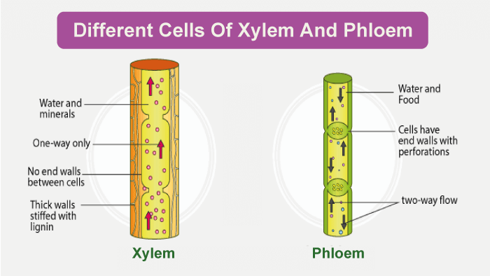
i) Xylem The main conducting tissue of vascular plants is called the xylem (Greek: xylos = wood). It is in charge of the conduction of inorganic and water-soluble solutes. Four distinct types of cells make up the xylem:
Along the principal axis of stems and roots, xylem tissue is arranged in a tube-like form. It is made up of tracheid, ray cells, fibers, vessels, and parenchyma cells. Tracheids are long tubes with separate cells open at both ends. There could be internal wall material bars that span the open area. Long tubes are created by joining these cells end to end. At maturity, tracheid and vessel members are dead. The secondary cell walls of the tracheid are thick and tapered at the ends. They lack the vessels' vessel-like end apertures. The ends overlap, and there are two pits at each end. Water can move between cells because of the pit pairs. While the majority of conduction in xylem tissue is vertical, rays aid lateral conduction along a stem's diameter. The vascular cambium gives birth to horizontal rows of parenchyma cells with a long lifespan, known as rays. ii) Phloem Phloem includes:
Phloem, which is a component of a plant's "plumbing system," is a tissue that is just as crucial. Phloem primarily transports dissolved food components throughout the plant. The partner cells and sieve-tube components that make up this conduction system lack additional walls. The vascular cambium's parent cells produce the xylem and phloem. Furthermore, fibers, parenchyma, and ray cells are often included. Members of sieve tubes put an end to end are used to create sieve tubes. Unlike vessel members in the xylem, the end walls lack apertures. Yet, the terminal walls are covered with tiny holes where cytoplasm travels from cell to cell. Sieve plates are the name for these permeable connectors. Sieve-tube members lack nuclei at maturity even though their cytoplasm is actively engaged in the conduction of dietary items. The partner cells that are tucked in between the sieve-tube components provide some kind of function, causing the conduction of food. The callus pad/callus, the colorless material that covers the sieve plate, comprises a callose polymer. This carbohydrate polymer is present in living sieve-tube members. Calcium stays in solution as long as the cell contents remain under pressure. As needed, food and other resources are moved up and down in plants via the phloem. Difference between Meristematic and Permanent Tissue
ConclusionThe cells that makeup tissues cooperate in carrying out certain tasks. Because of plants' complex way of existence, every tissue has been given a specific function. The plant tissues are divided into meristematic and permanent categories based on their capacity for division, function, and location. Meristematic tissues comprise similar, dividing, and differentiating cells supporting plant development. They are confined to certain areas like internodes, margins, and apices of roots and shoots. On the other hand, a mixture of several cell types leads to the creation of permanent tissues. These tissues comprise living or dead cells with massive vacuoles and strong cell walls. Their main job is to move the vital chemicals around within the plant's body. While the roles of the two tissues seem incompatible, they must work together and coordinate for the plant to grow and develop properly.
Next TopicDifference between
|
 For Videos Join Our Youtube Channel: Join Now
Feedback
- Send your Feedback to [email protected]
Help Others, Please Share


