Uses of Microscope
Introduction:
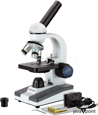
Microscopes are instruments used to magnify and observe microscopic objects or microscopic specimens; in other words, objects that are too small to be seen with the naked eye. They play a crucial role in various scientific and medical fields, enabling researchers and professionals to study the intricacies of microscopic structures that would otherwise be impossible to see by the naked eye. Microscopes have significantly contributed to advancements in biology, medicine, materials science, and many other disciplines. Microscopes have revolutionized scientific research by revealing the hidden world of microorganisms, cells, molecules, and nanoscale structures. They are indispensable tools for scientists, medical professionals, educators, and researchers who seek to understand the complexities of the microscopic realm.
There are several types of microscopes. Each of them plays a specific role and has different capabilities. The types are:
- Scanning Probe Microscopes: These microscopes use a physical probe to scan the surface of a specimen and create detailed images at the atomic level. Examples include Atomic Force Microscopes (AFM) and Scanning Tunneling Microscopes (STM).
- Confocal Microscopes: These microscopes use lasers and a pinhole to eliminate out-of-focus light, allowing for the creation of sharp, high-resolution images, usually used for thick specimens.
- Optical Microscopes: These are the most common type of microscopes and use visible light to magnify specimens. They can be further categorized into:
- Compound Microscopes: Utilize multiple lenses to magnify specimens, with the ability to achieve higher magnifications.
- Stereo Microscopes (or Dissecting Microscopes): Provide a three-dimensional view of larger specimens at lower magnifications, making them suitable for dissection and inspection.
- Electron Microscopes: These microscopes use electron beams instead of light, allowing for much higher magnifications and resolution. Electron microscopes include:
- Transmission Electron Microscopes (TEM): Transmit electrons through a thin specimen to create high-resolution images of internal structures.
- Scanning Electron Microscopes (SEM): Scan the surface of a specimen with electrons to generate detailed 3D images.
Working of a Microscope:
The functioning of a microscope involves multiple optical principles that work together to magnify and visualize small objects or specimens. Microscopes present a magnified image of a microscopic sample or object of interest, which otherwise would not be possible for the naked eye; this is the basic idea for the functioning of the microscope. The step-by-step functioning of a basic compound microscope is as follows:
- Illumination: The first step is the illumination of the specimen. The light source, which can either be a lamp fitted in the microscope itself or can just be a reflection of the light in the surrounding, provides light that passes through the condenser lens and is focused on the specimen.
- Formation of real image: The objective lens, which is the lens fitted in the microscope and is the closest to the object or the specimen. This objective lens converges the light reflected by the specimen, and the formation of a real and inverted image of the said specimen takes place. The image is formed between the oculus lens or the eyepiece, which is the lens through which the observer looks through the microscope, and the objective lens.
- Magnification: The microscope uses multiple lenses to magnify the image of the specimen. The eyepiece lens (ocular) is positioned close to the viewer's eye and typically magnifies the image further. The objective lenses, located near the specimen, provide the initial magnification. A typical compound microscope has several objective lenses with different magnification powers, which can be rotated into place as needed. The real image formed by the objective lens becomes the object for the eyepiece. The eyepiece further magnifies this image, allowing the viewer to see a greatly enlarged virtual image of the specimen.
- Final Image and Observation: The virtual image produced by the eyepiece is what the viewer observes through the eyepiece. This final magnified image is much larger than the actual size of the specimen, enabling the user to see fine details that would otherwise be invisible to the naked eye.
This is a basic outline of how a compound microscope works. Different microscopes have different functionalities.
History of Microscopes:
Microscopes are not a marvel of new ages. One of the earliest microscopes dates back to the 16th Century. A Dutchman by the name of Antoine Van Leeuwenhoek made one of the earliest microscopes, which consisted of a glass sphere in a metal frame. Leeuwenhoek's microscopes, though simple, had high magnification power and allowed him to make groundbreaking observations of microorganisms, earning him the title of "Father of Microbiology.". In 1590, another Dutchman, a spectacle maker, made what is said to be the first compound microscope.
It consisted of a convex and a concave lens that allowed for a relatively low level of magnification. Throughout the 18th and 19th centuries, numerous advancements were made in microscope design and optics, leading to improvements in resolution, magnification, and overall performance. This period saw the development of achromatic lenses, which significantly reduced chromatic aberration and improved image quality.
The 20th century witnessed the rise of electron microscopy, starting with the transmission electron microscope (TEM) in the 1930s and the scanning electron microscope (SEM) in the 1960s. These electron microscopes brought about a new era of high-resolution imaging, allowing scientists to study structures at the nanoscale level.
In recent times, advances in microscopy techniques continue to be made, including confocal microscopy, super-resolution microscopy, and various scanning probe microscopy techniques, each contributing to our understanding of the microscopic world in diverse scientific fields.
Uses of Microscopes:
Microscopes have a wide range of uses across various scientific, medical, industrial, and educational fields. Their ability to magnify and visualize tiny structures has revolutionized our understanding of the microscopic world. Some uses of a microscope are as follows:
1. Biology and Medicine:
The fields of biology and medicine are probably the most affected by microscopes. The ability to view microscopic bacteria, pathogens, and single-celled organisms made it easier to study their behavior and their basic functionalities, such as eating, digestion, reproduction, etc.
Microscopes also revolutionized the field of medicine as it became easier to observe how different microorganisms react to different chemicals. By observing these reactions, stopping the spread of these organisms or their eradication became easier. Some branches where microscopes are avidly used include
- Cell Biology: Microscopes are crucial for studying the structure and function of cells, including organelles, cellular processes, and cellular interactions. By studying the structure of cells, we can learn more about the functioning of not just single-celled organisms but also cell societies and more advanced organisms down to their cores.
- Histology: Microscopes are extensively used in medical settings for diagnosing diseases and conditions. Pathologists use microscopes to examine tissue samples (biopsies) for the presence of abnormal cells or pathogens, aiding in the diagnosis of cancer, infections, and other medical conditions.
- Microbiology: Microscopes are vital tools in microbiology for observing bacteria, viruses, fungi, and other microorganisms. Microbiologists use them to identify and study various pathogens, understand their structure, behavior, and life cycles, as well as to develop treatments and vaccines.
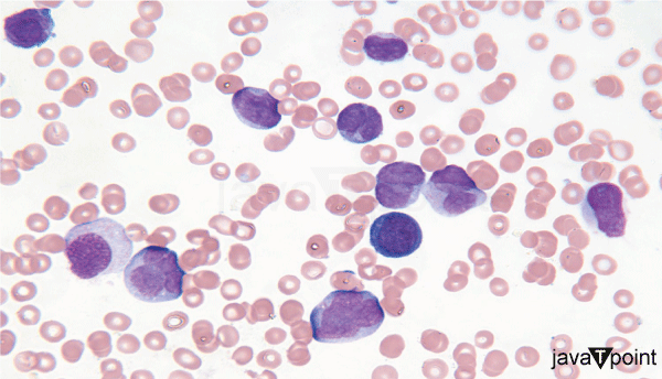
- Hematology: Microscopes are used in blood analysis to examine blood cells and identify abnormalities. Using microscopes, foreign bacteria, fungi, or parasites can easily be detected in the blood, which further helps in the eradication of pathogens.
- Pathology: Pathologists use microscopes to examine tissue samples (biopsies) to diagnose diseases like cancer.
2. Materials Science and Engineering:
In materials science and engineering, microscopes are used to examine the microstructure of materials, such as metals, polymers, ceramics, and composites. Scanning Electron Microscopes (SEMs) and Transmission Electron Microscopes (TEMs) help researchers visualize the arrangement of atoms and grains, study defects, and evaluate material properties.
- Metallurgy: Metallurgical microscopes are used to study the microstructure of metals and alloys, helping understand their properties and behaviors.
- Semiconductor Industry: Microscopes are used for quality control and failure analysis of semiconductor components.
- Nanotechnology: Scanning probe microscopes are used to visualize and manipulate nanoscale structures and particles. Nanoscale research relies heavily on electron microscopes like the Scanning Tunneling Microscope (STM) and Atomic Force Microscope (AFM).
3. Environmental Science:
Microscopes aid environmental scientists in analyzing soil, water, and air samples to study pollutants, microorganisms, and microplastics. Understanding these aspects is essential for environmental conservation and pollution control efforts.
- Microbial Ecology: Microscopes aid in studying the diversity and interactions of microorganisms in different environments. Using microscopes, microorganisms such as fungi, algae, bacteria etc can be studied in detail, and their interactions with one another can be understood properly.
- Water Quality Analysis: Microscopes are used to examine water samples for the presence of microorganisms and pollutants. Still or non-flowing water bodies such as lakes and ponds can be severely infected with fungi or other microorganisms, which can make them unfit for drinking or other human purposes. Microscopes make it easier to detect and differentiate these organisms.

4. Geology and Earth Sciences:
- Petrology: Microscopes are used to analyze rocks and minerals to understand their composition and formation.
- Paleontology: Microscopes aid in the examination of fossils to study ancient life forms. Traces of elements or other matter that could be crucial in discovering information about the fossil can be detected and analyzed easily with microscopes.
5. Forensics:
- Microscopes are used in forensic laboratories to analyze evidence like hair, fibers, and biological samples for criminal investigations.
- Using microscopes, smaller details can also be documented easily, which would otherwise be very difficult with the naked eye.
6. Education:
- Microscopes play a vital role in educational settings, allowing students to observe microscopic structures and better understand scientific concepts. Microscopes are also used in physics to learn about concepts of optics and the behavior of light.
- They are also essential for researchers across disciplines to observe and study intricate details that cannot be seen with the naked eye.
7. Quality Control and Research in Industry:
- In industries such as textiles, pharmaceuticals, and electronics, microscopes are used for quality control and research and development. They help inspect and assess the characteristics of products, ensuring they meet specific standards and specifications.
8. Art Conservation:
- Microscopes are used to study and analyze artwork to assess its condition and guide restoration efforts. Using microscopes, the quality, age, and type of canvas used can also be determined. All these are crucial information for determining the authenticity of the art piece.
9. Entomology:
- Entomologists use microscopes to study insects, their anatomy, behavior, and ecological research.
10. Genetics and Molecular Biology:
Microscopes are used in genetic research to visualize DNA, RNA, and proteins, as well as for techniques like fluorescence in situ hybridization (FISH). They are also used by students and scholars to observe DNA structures and proteins in biochemistry.
Conclusion:
It is an inarguable fact that microscopes have revolutionized not just the field of biology but rather other fields of science as well. The microscopes not only allow us to look at tiny objects but rather almost open up an entire microscopic realm for us to observe and study. A realm that otherwise is impossible to see with our eyes. In conclusion, microscopes have proven to be indispensable tools across a wide array of disciplines, revolutionizing scientific research and industry. From their foundational role in biology and medicine to their applications in nanotechnology and materials science, microscopes have allowed us to unlock the mysteries of the microscopic world.
|

 For Videos Join Our Youtube Channel: Join Now


