| |

Animal CellAnimal cells vary in size from several microns to several millimeters. The ostrich egg is the most substantial animal cell, occupying over 5.1 inches across and weighing about 1.4 kilograms. On the other hand, a neuron in the human body is of about 100 microns. The appearance of animal cells also diversifies. Some cells are flat, others oval or rod-shaped. These cells also have various shapes, like curved, spherical, etc. Most of the cells are so minute that they can be only can be seen under the microscope. Animal cells have a membrane around the nucleus that exhibits DNA; this is the reason it is also called eukaryotic cells. Animal Cell Structure:Animal cells are smaller than plant cells. The animal cells have an irregular shape because of the absence of a cell wall. Every animal cell is made up of the cell body and cell membrane. The cell is further divided into two parts, namely cytoplasm, and nucleus, which is surrounded by cytoplasm. The cytoplasm also contains many organelles, which we are going to discuss later. 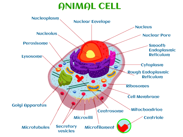
So, we can conclude that the structures of the animal cell mainly have the following three main components.
1. Cell membrane: Also called plasma membrane or plasma-lemma, the cell membrane is the outer protective sheath that separates. This membrane is semi-permeable, i.e., there can be a free exchange of various substances between the extracellular fluid (ECF) or outside of the cell, and the Intracellular fluid (ICF) inside of the cell. The thickness of the cell membrane varies from 80 Å to 112 Å. 2. Cytoplasm: The cell's cytoplasm is a semisolid (jelly-like) material containing about 75-85% water. The clear liquid portion of cytoplasm is called cytosol, and it also has various other particles of many shapes and sizes: proteins, carbohydrates, lipids, or electrolytes. The cytoplasm also contains many organelles that are of various structures and functions. 3. Nucleus: It is described later. Organelles in Cytoplasm:Cytoplasmic organelles are the cellular structures embedded in the cytoplasm. Organelles are acknowledged as the small organs of every cell. A plasma membrane binds some organelles, and many other organelles lack a plasma membrane. Every organelle has a particular structure and function. Let's talk about every organelle in detail.
MEMBRANE BUOND ORGANELLES1. ENDOPLASMIC RETICULUM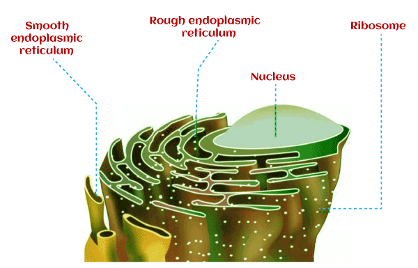
The endoplasmic reticulum is a membrane-bound organelle that is made up of a series of flat sacs in animal cells. It performs many functions, especially the synthesis, folding, transformation, and transport of proteins. The lumen of ER is filled with fluid that is called the endoplasmic matrix. The diameter of the lumen is about 395 to 695Å. Endoplasmic Reticulum Endoplasmic reticulum are of two types, namely
i) Rough endoplasmic reticulum (RER): The endoplasmic reticulum appears to be rough because of the presence of granular Ribosomes on its outer surface and that is the reason it is also called the granular endoplasmic reticulum and also sarcoplasm. Functions: The main function of RER is proteins synthesis. ii) Smooth endoplasmic reticulum (SER): The endoplasmic reticulum appears to be smooth because of the absence of granular Ribosomes on its outer surface and that is the reason it is also called the agranular endoplasmic reticulum and also ergastoplasm. Functions: The major functions include the synthesis of lipids and steroids. It also plays an important role in cellular metabolism like storage and metabolism of calcium, and also it helps in the detoxification of various other toxic substances. 2. GOLGI COMPLEX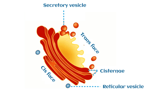
Discoverer Camillo Golgi, the Golgi body or Golgi complex, is an organelle involved in processing proteins. Generally, each cell has one Golgi complex, but some cells have more than one Golgi complex. These organelles are situated near the nucleus. It is made up of flattened sacs, various tubules and vesicles, and large vacuoles. Each Golgi complex has two ends or faces. The one where vesicles fuses are called cis face and where vesicles leave is called trans face. Functions: The main function of the Golgi complex is to deliver readymade proteins and lipids by processing, packaging, labelling. 3. LYSOSOMESDiscovered by de Duve, Lysosomes are organelles that are found all over the cytoplasm. They are also called suicidal bags as they contain hydrolytic enzymes. These enzymes are released from the Golgi complex at the M face or maturing face to degrade worn-out cells or organelles. Lysosomes are commonly found in WBCs, Liver, and Spleen. It is made up of a bilayer, which is why lysosomes have a very thick layer of membranes. Functions: Lysosomes are often referred to as the 'waste system' of a cell because of their degrading work. About 50 different hydrolytic enzymes, which are called acid hydroxylases are found in lysosomes. These enzymes help lysosomes in performing their function. 4. PEROXISOMESPeroxisomes were also discovered by de Duve that are also called microbodies. It is a single-layered membrane-bound organelle, unlike lysosomes that have bilayer membranes. Unlike lysosomes, peroxisomes are suppressed in the endoplasmic reticulum and not in the Golgi apparatus. Peroxisomes contain various enzymes like catalase, urate oxidase etc. Functions: It includes the breakdown of excess fatty acids and detoxification of harmful hydrogen peroxide and various other harmful metabolic products. It also provides oxygen utilization and acceleration of gluconeogenesis. It helps in the degradation of purine to uric acid and it also plays an important role in the formation of myelin and bile acids. 5. CENTROSOME AND CENTRIOLESThe centrosome is the membrane-bound organelle that contains two cylindrical structures that are made up of proteins and are called the centrioles. It is located almost in the centre of the cell that is close to the nucleus. Function: These organelles are responsible for the formation of spindle fibres and it also helps in the movement of chromosomes during cell division. 6. SECRETORY VESICLESAs the name suggests, these organelles contain secretory substances. These organelles are present all over the cytoplasm. These organelles are produced in the endoplasmic reticulum after passing trans face. Later, they are packed and processed in the Golgi complex and are ruptured when required to release the substance into the cytoplasm. 7. MITOCHONDRION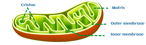
Mitochondria are oval and rod-shaped membrane-bound organelles that are bound by a double-layered membrane. The outer membrane contains enzymes like Acetyl-CoA synthetase and on the other hand, the inner membrane is folded into multiple projections called cristae, which contains exosomes, and the middle part is called the matrix part that is the site of the Krebs cycle. These organelles are also called semi-autonomous cell organelles as they contain their own DNA. The number of mitochondria in a particular cell depends on the metabolic activity of the cell, the more the activity of the cell more will be the mitochondria in number. Functions: The main function of the mitochondria is ATP synthesis. It also has other functions like storage of calcium and detoxification of ammonia. 8. NUCLEUSDiscovered by Robert Brown, the nucleus is also called the cell's control centre, and its study is called Karyology. It is the largest cellular organelle that has a diameter of 12 µ to 20 µ. These organelles are although present in all animal cells but are absent in the red blood cells. Depending upon the number of nuclei present in the cell, the cell can be termed as uninucleated (single nucleus) and multinucleated (multiple nuclei). 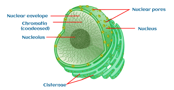
Structure: It consists of:
i) Nuclear Membrane It is a double-layered membrane that encloses nucleoplasm and the space between the two layers is known as perinuclear space. Some pores are present in the membrane called nuclear pores that breaks the continuity of the nuclear membrane. It is the site where nucleoplasm and cytoplasm exchange materials. ii) Nucleoplasm Nucleoplasm is also called nuclear sap that consists of nucleic acids, enzymes, etc. It is the site of DNA, RNA, ribosomal subunits. This organelle surrounds the chromatin and nucleolus. The nucleoplasm also contains the nuclear hyaloplasm, which is the soluble fraction of nucleoplasm. iii) Chromatin Chromatin is a thread-like chromosomal material that becomes visible during the interphase. Chromatin is made up of large DNA molecules. Chromatin consists of DNA cells with histone and non-histone proteins, which is why chromatin is also called the DNA-histone complex. iv) Chromosomes The chromosome is the supercoiled form of chromatin. It contains the complete blueprint genome and is packed with a number of genes. It becomes visible only at the time of cell division. The different organism has a different set of chromosomes. For example, if we talk about the human body, it has 23 pairs of chromosomes in somatic cells and 23 single sets in sex cells. Functions: The nucleus controls cell metabolism, protein synthesis, RNA synthesis, and it is also the site of the formation of subunits of ribosomes. NON- MEMBRANE BOND ORGANELLES:1. RIBOSOMESDiscovered by Palade, Ribosomes are non-membranous cell organelles that are found in both prokaryotic and eukaryotic cells. In Prokaryotic cells, they occur freely in the cytoplasm, but in Eukaryotic cells (Example- Animal Cell), they are also found attached or inside the Endoplasmic Reticulum (ER). It consists of 40% of proteins and 60% of ribonucleic acid (RNA). These organelles are of two types- 70S and 80S. 80 S type of ribosomes is found in animal cells. The 80S ribosome is made up of two subunits that are 60S, which is the larger subunit and 40S, which is the smaller subunit. Function: The main role of ribosomes is the synthesis of proteins. The attached ribosomes are mainly responsible for synthesising enzymatic proteins, hormonal proteins etc., while free ribosomes produce proteins in haemoglobin. 2. CYTOSKELETONThe cytoskeleton is the cellular organelle made up of the network of fibres crisscrossing through the cell wall that helps maintain the shape of the cell. Cytoskeleton fibres are made up of 3 important parts:
i) Microtubule 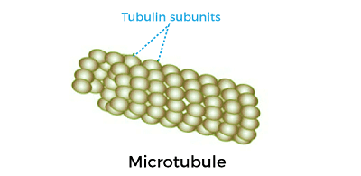
Microtubules are generally 25 nm in diameter that helps in determining the shape and structural strength of the cell. ii) Intermediate filaments 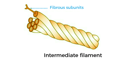
Intermediate filaments are generally 10 nm in diameter that helps to maintain the shape of the cell. iii) Microfilaments 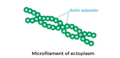
Microfilaments are generally 5 nm in diameter that gives strength and provide resistance to the cell against the pulling forces.
Next TopicBudding
|
 For Videos Join Our Youtube Channel: Join Now
Feedback
- Send your Feedback to [email protected]
Help Others, Please Share


