| |

Ventricles of the BrainOur brain controls a variety of cognitive and physiological functions. The ventricles are a network of connected chambers that are essential for the circulation of cerebral spinal fluid (CSF) and are at the centre of its operation. The first ventriculography was carried out on an ox's brain by renowned artist Leonardo da Vinci, who also sketched the Mona Lisa. Francis Jean Magendie later confirmed Domenico Felice Antonio Cotugno's description of the relationship between the cerebral ventricles and subarachnoid space from the 17th century. Overview of Ventricles: The brain's ventricles are an intricate system of interconnecting chambers. The ventricular system consists of 2 lateral ventricles, the third ventricle, the cerebral aqueduct and the fourth ventricle. The lateral ventricles communicate with the third ventricle through interventricular foramen, and the third ventricles communicate with the fourth ventricle through the cerebral aqueduct. Structure and Composition: Ependymal cells, a specialised layer of cells, line the ventricles. These cells create and control CSF, a transparent liquid that covers and supports the brain. The CSF acts as a barrier, providing nutrients to the brain tissue and absorbing shocks. The CSF can readily flow between the ventricles since they are connected to one another. Functions of Ventricles:
Important Functions of CSF<
Types of Ventricles in the BrainTypes of ventricles in the brain, their roles, and significance are listed below: 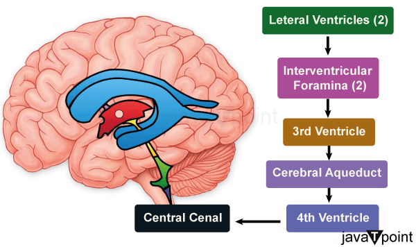
Lateral Ventricles:The lateral ventricles are the largest and most prominent among the ventricles. There are two lateral ventricles in the brain. They are located close to the corpus callosum, deep inside the cerebral hemispheres. A small opening known as the interventricular foramen connects these ventricles. The cerebrospinal fluid (CSF), a transparent fluid that protects and cushions the brain and spinal cord, is created by the lateral ventricles. The exchange of nutrients and waste products between the brain and blood arteries is also aided by CSF. These ventricles also play a role in controlling brain temperature. Third Ventricle:The third ventricle is a median slit-shaped cavity that is found between the two thalami and a portion of the hypothalamus, which is situated in the midline of the brain underneath the lateral ventricles. The interventricular foramen connects this tiny, slit-like cavity to the lateral ventricles. Through the cerebral aqueduct, the third ventricle communicates with the fourth ventricle. Through its connection to the hypothalamus, the third ventricle regulates hormone secretion, which is essential for maintaining homeostasis. Additionally, it takes a role in the regulation of autonomic processes like hunger, thirst, and sleep. The third ventricle also participates in the movement and circulation of CSF. Fourth Ventricle:In front of the cerebellum, at the base of the brainstem, is where the fourth ventricle is located. It resembles a diamond-shaped chamber and is connected to the spinal cord's central canal. Through the cerebral aqueduct, the fourth ventricle communicates with the third ventricle. The modulation of motor coordination and control depends on this ventricle. Through its connections to the cerebellum, it also plays a significant part in maintaining balance and posture. The fourth ventricle acts as a passageway for CSF to move throughout the brain and spinal cord, assisting in the elimination of waste materials. The cerebral aqueduct, also called the Sylvius' aqueduct, is a tiny passageway that links the third and fourth ventricles of the brain. It serves as a conduit for the flow of CSF as it passes through the midbrain. White matter surrounds the cerebral aqueduct, which is essential for maintaining normal CSF flow throughout the brain. Cisterns ConceptCisterns are anatomical structures found within the central nervous system that play a crucial role in the circulation and distribution of cerebrospinal fluid (CSF). These fluid-filled spaces are located in various regions of the brain and spinal cord, aiding in the protection and nourishment of these vital structures. Let's explore cisterns in more detail. Cisterns are expanded spaces that are located in the subarachnoid space, which is a region between the arachnoid and pia mater, two of the three membranes that protect the brain and spinal cord. Cisterns come into the role when CSF spreads out and gets collected in particular areas, forming reservoirs that promote the exchange and movement of this fluid. The cisterna magna, also known as the cerebellomedullary cistern, is one of the biggest and most well-known cisterns. Between the cerebellum and the medulla oblongata, this cistern is situated at the back of the brain. It acts as a reservoir for the fluid, enabling its passage between the brain and spinal cord, and plays a crucial function in the circulation and absorption of CSF. The pontine cistern, which is located around the pons n the ventral portion of the brainstem, is another significant cistern. The CSF in this cistern protects the sensitive brainstem regions by acting as a cushion. Critical structures like the basilar artery and oculomotor nerve are crucial for protecting environments in the interpeduncular cistern, which is situated at the base of the brain between the two cerebral peduncles. Cisterns are found throughout the spinal cord in addition to the brain. For instance, the lumbar cistern is situated below the spinal cord's end in the lower spinal canal. The extraction of CSF during a lumbar puncture or spinal tap frequently takes place in this area since it offers a reasonably safe and accessible location for the procedure.
Next TopicBlood Brain Barrier
|
 For Videos Join Our Youtube Channel: Join Now
Feedback
- Send your Feedback to [email protected]
Help Others, Please Share


