Brain CT Scan
Modern healthcare has been revolutionised by the advent of medical imaging technology, which allows medical practitioners to accurately diagnose and treat a variety of illnesses. The Computed Tomography (CT) scan is an example of a cutting-edge technology that is used to diagnose illnesses, direct medical interventions, and track the effectiveness of treatment.
Fundamentals of CT Scans
A CAT (Computerised Axial Tomography) scan, commonly referred to as a CT scan, is a non-invasive medical imaging procedure that produces fine-grained cross-sectional images of the body's internal components. In contrast to conventional X-rays, which provide 2D images, CT scans use a number of X-ray beams and specialised detectors to produce 3D images. These cross-sectional pictures, or "slices," give a thorough perspective of the blood arteries, bones, tissues, and organs of the body.
Principles of CT Scans
The fundamental idea behind CT scanning is the scattering of X-rays as they travel through various body components. Depending on the density of the tissue, X-rays, a type of electromagnetic radiation, can enter it to various degrees. Bones and other dense structures absorb more X-rays, which is why they appear white in CT pictures. Contrarily, less dense objects, such as organs and soft tissues, permit more X-rays to flow through, giving the images a darker tone.
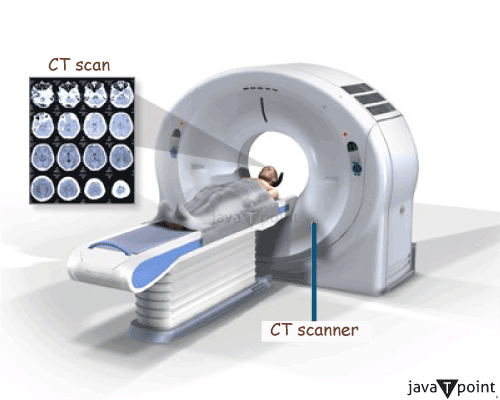
A gantry, an X-ray tube, and a number of detectors make up a CT scanner. During the scanning procedure, the gantry, which houses the X-ray tube and detectors, revolves around the patient. A computer processes the numerous X-ray beams that are released and detected from different angles as the gantry spins in order to rebuild intricate cross-sectional images.
Applications of CT Scans
CT scans have a broad spectrum of applications across various medical disciplines:
- Diagnosing Diseases: A variety of medical disorders, such as tumours, infections, fractures, and internal bleeding, can be detected and diagnosed using CT scans.
- Guiding Medical Procedures: In order to ensure exact targeting and lower the risk of complications, doctors frequently employ CT scans to guide biopsies, aspirations, and other interventional operations.
- Trauma and Emergency Care: CT scans are essential in evaluating injuries swiftly in situations involving trauma or sudden emergency, enabling medical professionals to act promptly and accurately.
- Cancer Staging: CT scans are essential for cancer staging because they allow physicians to assess the degree of tumour spread and develop the most effective treatment plans.
- Neuroimaging: CT scans are used in neuroimaging to identify neurological conditions such as strokes, brain tumours, and intracranial haemorrhage, allowing for quick diagnosis and effective treatment.
Benefits of CT Scans
CT scans offer several advantages over other imaging modalities:
- Speed and effectiveness: CT scans are suitable for emergency circumstances since they are reasonably quick, frequently taking only a few minutes to complete a scan.
- High-resolution Imaging: CT scans offer a high level of clarity and detail, making it possible to see microscopic structures and subtle abnormalities.
- Versatility: CT scans are appropriate for a wide range of medical disorders since they can image different body regions.
- Non-invasive: CT scans are non-invasive since they often don't need to be performed with surgical incisions or the injection of contrast material.
- Painless: The average CT scan patient feels little to no pain, which improves patient comfort.
Limitations and Considerations
There are certain limitations and considerations to be kept in mind:
- Radiation Exposure: Ionising radiation is used in CT scans, which has the potential to be dangerous, particularly with repeated or cumulative exposures. Before recommending CT scans, doctors carefully assess the advantages vs the hazards.
- Allergies to contrast material: Some patients may experience negative effects if they are allergic to the contrast material used in CT scans.
- Cost: Compared to certain other imaging modalities, CT scans can be somewhat pricey, which patients and healthcare professionals may want to take into account.
- Particular Populations: Because of radiation concerns, pregnant women and young patients need to be treated with extra caution.
Brain CT Scan Looks Like
An accurate cross-sectional image of the brain is produced by a brain CT scan, which enables medical specialists to see the organ's interior structures and identify problems. The images are in grayscale, with various shades of grey standing for various brain tissue densities.
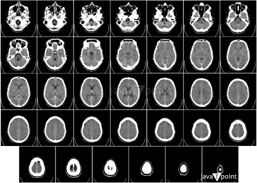
- Bone structure: The skull appears as a bright white outline in a brain CT scan because bones are dense structures that significantly attenuate X-rays. The skull is a wonderful point of comparison when analysing the architecture of the brain because it is simple to see its shape and structure.
- Brain Tissue: The actual brain tissue takes on a variety of shades of grey depending on its density. Compared to white matter, which contains nerve fibres with myelin sheaths, grey matter, which contains nerve cell bodies, is clearly lighter. Because it is less dense and allows X-rays to pass through it readily, the CSF that covers and protects the brain appears black.
- Ventricles: The ventricles of the brain are liquid-filled chambers that facilitate the passage of cerebrospinal fluid. These show up as dark, black areas on the CT scan because of the low fluid density.
- Gyri and Sulci: The gyri are the raised folds on the surface of the brain, and the sulci are the grooves or furrows found between them. These traits contribute to the peculiar appearance of the brain's convoluted surface and can be noticed on a brain CT scan.
- Blood Vessels: A CT scan may reveal large blood arteries, particularly those that are close to the surface of the brain. The contrast with the surrounding brain tissue makes these look like thin, white lines.
- Abnormalities: A brain CT scan can identify a variety of anomalies, including tumours, haemorrhagic lesions, infarcts, and signs of trauma or damage.
What happens During the Brain CT Scan
Preparation and Patient Information:
The patient is usually requested to change into a hospital gown and take off any metal objects, such as jewellery and hairpins, before the CT scan because they can interfere with the imaging procedure. The technologist or radiographer will enquire about the patient's medical background, such as any allergies, prior operations, or underlying medical disorders. If a patient is pregnant, it is crucial to let the medical personnel know since extra care could be required.
- Positioning on the CT Table: The patient is laying on a small CT examination table that slides into the scanner's circular entrance. To get precise and clear scan results, one must remain motionless the entire time.
- Intravenous Contrast Injection (Optional): On rare occasions, the use of a contrast dye can enhance the visibility of particular brain structures or illnesses. If intravenous (IV) contrast is required, a small IV line is inserted into a vein, usually in the arm. Prior to the scan, the contrast agent is then injected into the bloodstream to highlight blood vessels and abnormal tissue and provide better diagnostic data.
- CT Scan Process: Once the patient is lying comfortably, the CT scan can begin. As the CT scanner revolves an X-ray tube around the patient's head, a detector keeps track of the X-rays that pass through the patient's brain. Fine cross-sectional images of the brain are created by the detector and X-ray tube working together. These images are also known as slices or tomographic images. Using these images, which were collected from different angles, a complete 3D model of the brain is produced.
- Monitoring and Communication: From a nearby control room, the tech continuously monitors the patient during the CT scan. They may speak to the patient over an intercom system to provide instructions or reassurance during the surgery. The patient is required to remain composed and relaxed throughout the scan.
- Scanning Duration: Although the total scanning time can vary depending on the complexity of the scan and the usage of contrast, the actual scanning procedure frequently only takes a few minutes. It may be necessary to perform numerous scans to get a complete picture of the brain in some cases.
- Post-Scan: Following the completion of the brain CT scan, the patient is free to leave the examination table. If contrast dye was employed, the IV line might be removed, and the patient would be closely monitored for any allergic reactions or negative side effects. Immediately following surgery, patients often resume their normal activities.
- Image Analysis and Diagnosis: A radiologist, a specialist physician with training in the interpretation of medical pictures, processes the CT images that have been obtained. To find any anomalies, like tumours, haemorrhages, or other diseases affecting the brain, the radiologist examines the images. To help with future diagnosis and treatment planning, they will write up a thorough report that is shared with the patient's primary care physician or another expert.
Precautions
Here are some important precautions to keep in mind:
- Inform the Healthcare Provider: Before the brain scan, it is very important to inform the doctor of any current medical conditions, allergies, or previous operations. Any metal implants, devices, or health conditions that they are aware of may have an impact on the type of scan or the use of contrast material.
- Pregnancy: It is important to let the healthcare professional know if pregnant. A growing foetus may be at risk from radiation exposure during a CT scan, and pregnant women may also need to take extra precautions when having some MRI scans.
- Metal and Electronics: An MRI or CT of the brain uses strong magnetic fields or X-rays, so it is crucial to take out any metal objects from the area of the body being scanned. Detachable dental work, eyeglasses, hearing aids, jewellery, and hairpins should all be removed. When undergoing an MRI, mobile phones and smartwatches should be kept outside the scanning room due to the strong magnetic field's potential to damage them and impair their functionality.
- Contrast Dye Allergy: If a contrast agent or iodine allergy has ever been affected, inform the medical professional before having a brain scan that uses contrast dye. Despite somewhat rarity, allergic responses to contrast material are a possibility, thus vigilance is advised.
- Claustrophobia (for MRI): Inform the doctor if having claustrophobia or a fear of cramped places before undergoing an MRI. They might advise using an open MRI scanner or learning relaxation techniques as means of controlling your anxiety throughout the procedure.
- Kidney Function: Some persons with kidney problems may be at risk from the contrast substance used in CT scans. Tell the doctor right once if someone has a history of renal disease or any issues with kidney function.
- Medication and Medical History: Make sure the doctor is aware of all the medications one is taking, including prescription, over-the-counter, and dietary supplements. Before the scan, some medications, especially those that interact with the contrast agent, may need to be changed or temporarily stopped.
- Nursing Mothers: The potential hazards and advantages of the brain scan for nursing mothers must be thoroughly discussed with the medical professional. Depending on the type of scan and the application of contrast dye, they can advise on whether breastfeeding should be stopped.
- Follow Pre-scan Instructions: Before the brain scan, the doctor will give specific instructions, such as fasting or dietary restrictions, especially if a contrast agent is going to be used. To ensure the accuracy and safety of the scan, it is crucial that one must carefully follow these directions.
Are Brain CT Scans Safe?
In general, CT scans are thought to be secure and non-invasive, offering patients little discomfort while still giving crucial diagnostic data. However, there could be hazards involved with CT scans, just like with other medical procedures using radiation. The main issue is ionising radiation exposure, which over time can harm cells and raise the risk of developing cancer. Although CT scans employ a relatively low amount of radiation, repeated or unneeded scans can lead to an accumulation of radiation exposure, especially in populations that are more susceptible, such as children and young adults.
When doing CT scans, medical personnel adhere to the ALARA (As Low As Reasonably Achievable) approach to reduce these hazards. This means that they work to use the least amount of radiation feasible to gather diagnostic data. The development of low-dose CT protocols and iterative reconstruction techniques, which further reduce radiation exposure without sacrificing image quality, is also a result of technological improvements.
The advantages of getting a head CT scan when medically necessary greatly outweigh any potential dangers for the vast majority of people. Brain tumours, strokes, traumatic brain injuries, and bleeding inside the brain can all be diagnosed and monitored with the help of CT scans. They enable quick assessment of vital conditions in emergency situations, which can save lives.
The need for a CT scan must be carefully assessed by medical professionals, who must also proceed with prudence. When alternate imaging techniques, like magnetic resonance imaging (MRI), maybe just as useful, they must balance the potential benefits against the radiation hazards. CT scans are still essential for prompt diagnosis and adequate treatment planning in cases when MRI is not feasible or easily accessible.
Medical personnel should follow established norms and regulations while using CT scans to protect patient safety. Due to the potential hazards to the growing foetus, they should acquire informed consent from patients, especially when thinking about repeat scans or when imaging pregnant people.
|

 For Videos Join Our Youtube Channel: Join Now


