| |

Chromosome: definition, structure, types, & compositionA chromosome is a thread-like structure located in the nucleus of cells such as plant, animal and human cells. Each chromosome is made of a molecule of DNA (Deoxyribonucleic acid) and histone proteins. Its unique structure keeps the two DNA strands tightly wrapped around the histone proteins. So, DNA is packaged into a chromosome or a chromosome is a bundle of tightly-coiled DNA. 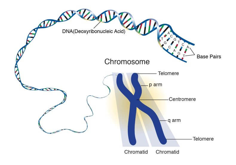
Organization of chromosome:DNA is wrapped multiple times around histone proteins, which is a histone octamer as it is made of eight histone molecules four above and four below. The structure formed due to the wrapping of DNA around histone protein is called nucleosomes. The nucleosomes are densely packed to form chromatin which coils up tightly to make chromatin loops. These loops further wrap around each other and thus chromatin becomes highly folded to make a full chromosome. 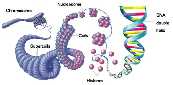
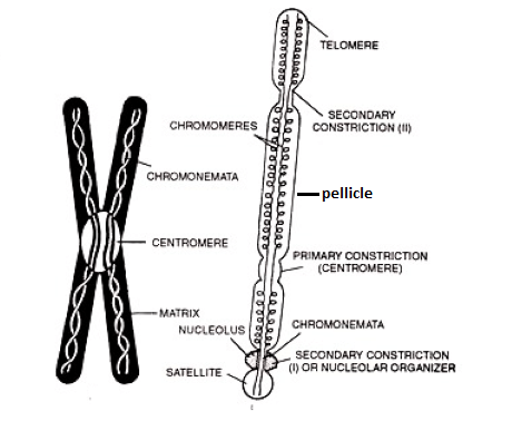
The DNA wrapped around the histone proteins has 147 base pairs. We can say that a histone octamer holds the 147 base pairs. Nucleosomes look like beads of a necklace. The DNA between two nucleosomes is called linker DNA and it has around 53 base pairs. So one nucleosome including its linker DNA contains 200 base pairs. The name chromosome is derived from two Greek words chroma which means colour and soma which means body. It got this name as it was the only cell structure that was deeply stained by colourful dyes used in by the scientists in their research. The chromosomes are usually not visible even under a microscope when the cell is not dividing. It is visible under a microscope only when the DNA in a chromosome gets tightly packed during the metaphase stage of cell division. Discovery of Chromosomes:In 1842, Karl Nageli, was the first person who had seen rod-like structures present in the plant cells' nucleus. It is generally known that chromosomes were first discovered by Walther Flemming in 1882. Later, in 1888, von Waldeyer-Hartz coined the term 'chromosome'. In 1902, two scientists Theodor Boveri and Walter Sutton independently found that chromosomes are the carrier of genetic material or genes. Besides this, the shape of chromosomes varies from species to species. For example, bacteria generally have circular chromosomes. Whereas, plants, animals, and humans have linear chromosomes in their cells. The number of chromosomes also varies from species to species. However, its number is the same for all the cells of a given species. For example, all human cells contain 23 pairs of chromosomes or 46 chromosomes arranged in 23 pairs; 22 pairs of autosomes and one pair of sex chromosomes (XX for females and XY for males). The fruit fly has four pairs of chromosomes, rice plant has 12 pairs of chromosomes and a dog has 39 pairs of chromosomes. So, every species has its own characteristic number of chromosomes. The cells that make the body of the organism but do not take part in forming new a organism during reproduction are called somatic cells. All human cells are somatic cells except for sex cells such as sperm and egg cell. Somatic cells are diploid (2n) as they contain 23 pairs of chromosomes; two copies of each chromosome. However, only the gametes or sex cells such as human sperm and egg are haploid (1n) as they have one copy of each chromosome (only one chromosome of a pair); a total of 23 chromosomes. So, they are called haploid (1n) cells. The two gametes fuse or unite to form a single diploid (2n) cell that contains two copies of each chromosome; 23 pairs or 46 chromosomes. Thus, each parent provides one chromosome to make a pair so the children get half of their chromosome from their father and a half from their mother. Thus, one set of chromosome comes from father and second set comes from the mother. The major function of Chromosomes is to provide stability to DNA and to ensure that the DNA is replicated and distributed equally during cell division. Structure of Chromosome I Main Parts of a Chromosome:A chromosome is made of histone proteins and DNA strands that form a 3-dimensional (3D) structure called a double helix. A chromosome generally has eight parts.
Types of chromosomes I Shapes of chromosomesAll the 46 chromosomes in a human cell do not look the same. They are not exactly like one another. They are of different types based on their shape, structure, and function. i) Chromosome types based on the number of centromeres present on a chromosome:
ii) Chromosomes types based on the location of the centromere on a chromosome: 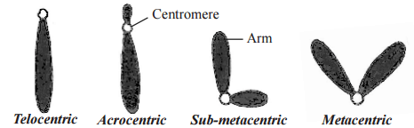
iii) Types of Chromosomes based on their function:
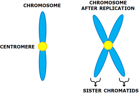
How many DNA strands in a chromosome?Before replication, when the cell is not dividing, a single chromosome has one DNA molecule (double-stranded DNA), after DNA replication, a chromosome has two DNA molecules (two double-stranded DNA). 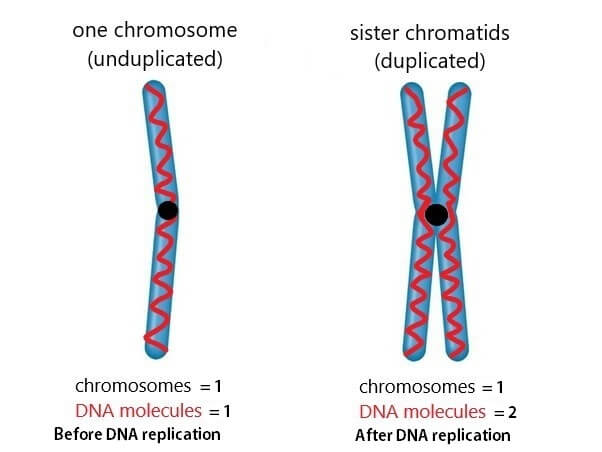
We can say that amount of DNA increases after replication but there is no increase in the number of chromosomes. The identical copies that form the two halves of a replicated DNA are called sister chromatids. In the later stages of cell division, the sister chromatids get separated longitudinally to give rise to individual chromosomes. One chromatid consists of one DNA helix (two spirally twisted or interconnected strands of DNA). So, 2 chromatids = 2 helixes = 4 DNA strands. So, how many strands a chromosome can have depends on the stage of the cell cycle. Chemical composition of chromosome:It is made of three components, DNA, proteins and RNA in small amounts. The amount of DNA ranges from 30 to 40 %, the amount of proteins from 50 to 65%, and small amount of RNA from 1 to 10% that is formed during DNA transcription. However, this chemical composition may vary from organism to organism. The protein present in DNA is of two types histone and non-histones proteins. The histone protein is present in large amount (90% of total protein), whereas non-histone is present in small amount (around 10% of total protein). The histone protein is highly basic, whereas, the DNA is acidic. So, basicity is neutralized by acidity as a result it gives rise to a neutral chromosome which has no acidity or basicity. Besides this the DNA is present in two forms in chromosomes; Euchromatin and Heterochromatin. Euchromatin means true chromatin as it contains genes that are functional or who code for protein synthesis. Heterochromatin means false chromatin. As the name suggests, it does not contain functional genes capable of coding for protein synthesis. Euchromatin is lightly stained DNA as it is a little loose so that all the process related to protein synthesis can take place easily. Whereas, heterochromatin is darkly stained DNA as it is densely packed. Study of Chromosome:What is karyotyping?Karyotyping is a technique which is used to study the chromosomes of a species. In this technique, chromosomes are isolated, stained and then photographed to create a micrograph image of chromosomes arranged in pairs. It also helps find out abnormalities in chromosomes. What is a karyotype?It is a picture of all the chromosomes of a person. It shows all chromosomes found in a cell. It describes the chromosomes count and shows their characteristics such as length of the chromosome, the position of the centromere, banding pattern, etc. What is Karyogram?It is also known as the Idiogram. It is the diagrammatic representation of the real stained chromosomes in a standard format in which chromosomes are arranged in an appropriate order based on their size, length, the position of chromosomes, degree of distribution of heterochromatin region, etc. such as in the order of decreasing size. It is a standard format of chromosomes that is created by the rearrangement of the photomicrographs of chromosomes. The chromosomes are photographed then each chromosome is cut and pasted to create karyogram or ideogram. The homologous chromosomes are kept side by side and then arranged by decreasing length.
Next TopicGiant Chromosomes
|
 For Videos Join Our Youtube Channel: Join Now
Feedback
- Send your Feedback to [email protected]
Help Others, Please Share


