| |

Golgi body | Golgi apparatus | Golgi Complex | Cell organelleGolgi body is one of the important cell organelles. It is also known as Golgi apparatus or Golgi complex. Golgi body was discovered by Camillo Gogi in 1989 in the cells of cats and barn owl. However, its ultra-structure is given by Dalton, so, sometimes it is also known as Dalton's complex. It is also called lipochondria. Besides this, in plant cells, it is known as Dictyosome. It is found in all eukaryotic cells, except mature mammalian red blood cells (RBCs). It is also absent in prokaryotic cells as prokaryotes lack membrane-bound cell organelles. Size of Golgi body:Its size varies according to the functions of cells, for example, it is bigger in secretory cells, the cells that have a secretory function such as the liver cells that have to secrete various secretions. In non-secretory cells, it is comparatively smaller in size such as in muscle cells. Number of Golgi body:There is only one Golgi body per cell in animal cells. However, in plant cells, the number of Golgi bodies is not fixed and varies from cell to cell, e.g., there can be 6 to 12 Golgi bodies in a plant cell. The plant cell with more secretory functions will have more of it. In animals or vertebrates, the Golgi body is perinuclear or close to the nucleus. Whereas, in invertebrates and plant cells, it is scattered in the cytoplasm. Furthermore, there is also a clear cytoplasmic region around this cell organelle, which is called zone of exclusion as it does not contain any organelle. Furthermore, Golgi body is a pleomorphic organelle, which means it can change its shape as per the functions performed by it. Structure of Golgi body:It is made up of stacks of membrane-bound flattened sacs called cisternae and vesicles and vacuoles. It has two faces; a cis face (receiving face) that receives proteins and lipids and a trans face (supplying face) that supplies modified or processed proteins to their different destinations. Its main parts are described below; 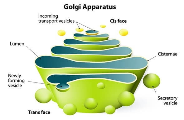
i) Cisternae: They are flattened sac like structures which are not straight. They are slightly curved and their ends are swollen. These swollen ends are known as Golgian vacuoles. The thickness of each cisterna ranges from 15 to 20 nm and the gap between two adjacent cisternae is around 20 to 30 nm. The number of cisternae present in a Golgi body is also variable. They can be 6-12 in number in animal cells and 10 to 20 in plant cells. However, in fungi, there is only one cisterna so, in fungi, it is also called a unicisternal Golgi body. The cisternae at the cis region differ from the cisternae of the trans region in shape, size, enzymatic activity, content, and vesicles. The cisternae at cis region or face are towards the rough ER. Whereas, the cisternae at the trans region or face are towards the plasma membrane. The cisternae located between the cis and trans regions are called medial cisterns. The cis face cisternae, medial cisternae and trans face cisternae contain different enzymes that are required to modify, sort, and pack proteins before their transportation. ii) Vesicles: They are also spherical structures but they are smaller than vacuoles and they are located towards the convex side or cis face of the Golgi body. iii) Vacuoles: They are spherical structures with a diameter of around 600 Angstrom. They are usually found close to the concave side or trans face of the Golgi body. Besides this, Golgi body shows polarity as it has two sides; one is concave and the other is convex side. The concave side, which is towards the plasma membrane, is known as the Trans face or maturation face. Whereas, the convex side, which is towards the nuclear membrane, is known as the cis face or formative face. How Golgi body works?A part of the membrane of rough ER buds off and forms a transport vesicles to transport protein to Golgi body. The transports vesicle travels towards the cis face of the Golgi body and then fuses with the cisterna at the cis face and releases its content (proteins) into the cisterna. The vesicles coming from RER contain proteins, while, the vesicles coming from SER contain lipids. Golgi body modifies the protein and lipid products. The modification occurs in the middle cisternae. The modified proteins move from one cisterna to adjacent cisterna again through transfer vesicle that buds off from swollen edge (Golgian vacuole) of a cisterna. The golgian vacuole increases in size and buds off to form vesicles containing the final modified product. These vesicles may contain hydrolytic enzymes, so in that case, they are called lysosomes. The modified products through vesicles reach their destinations such as other organelles, cytosol, or outside the cells. The vesicles which are like small bubbles and transport proteins from ER to GA and from GA to their destinations are generated from the membrane of ER and GA through budding. When vesicles reach their destination their membrane is fused to release the protein cargos. So, the entry or cis face receives the proteins from the ER and modifies these proteins. In the medial region, the sugar is added to proteins and lipids to form glycoproteins and glycolipids respectively. At exit or trans face, final processing of proteins occur that involves sorting and packaging and then transport of proteins to their destination through vesicles. Some of the processed proteins are transferred through secretory vesicles from trans face to plasma membrane from where they are discharged into the extracellular fluid through exocytosis. Whereas, some processed proteins are transferred to the plasma membrane by vesicles to integrate or to be used in the plasma membrane. Some proteins leave the trans face in storage vesicles such as lysosome. So, we can say that lysosome is formed from the Golgi body. Besides this, the Golgi body tags the proteins with sugar molecules that work as a shipping label for the protein molecules. So, its main job is modification, packaging and transport of proteins and synthesis of the plasma membrane and lysosomes. Cytochemistry of Golgi body:Golgi body contains lipids as it shows affinity to silver and osmium stains. Its membrane is made of equal parts of proteins and lipids and it also contains carbohydrates. A concentration gradient of polysaccharide is also found across this organelle. The higher concentration is at the maturating or trans face and the low concentration is at the formative or cis face. Besides this, it also contains fatty acids, vitamin C and carotenoids and various enzymes such as phosphatase, glycosyltransferase, mannosidases, phospholipases, oxidoreductases, transferases, and kinases. Origin of Golgi body:There are many theories for the origin of the Golgi body. However, as per the electron microscopic studies, it was found that vesicles that arise from the outer nuclear membrane or ER form the Golgi body. These vesicles are called transition vesicles. They may form Golgi complex or may fuse with any existing cisterna membrane present in the cytoplasm. Functions of Golgi complex:
So, we can say that the main function of GA is secretory. It produces secretory vesicles and vacuoles that contain secretions of cells like proteins, enzymes, etc. Besides this, it also takes part in the formation of the cell wall, plasma membrane and lysosomes.
Next TopicMitochondria
|
 For Videos Join Our Youtube Channel: Join Now
Feedback
- Send your Feedback to [email protected]
Help Others, Please Share


