| |

Layers of BrainThe three membrane layers surrounding the brain and spinal cord are referred to as the meninges, with the part surrounding the brain being known as the cranial meninges. The three layers are the dura, arachnoid, and pia; the Latin word "mater," which means "mother," is sometimes used after these names (e.g., dura mater, arachnoid mater, pia mater). The dura, which translates from Latin as "hard," is a layer of thick connective tissue that attaches to the inside surface of the vertebrae and skull. The extremely thin, transparent membrane known as the pia, which is directly adhered to the surface of the brain and spinal cord, is superficial to the arachnoid. This thin, wispy membrane lies only deep to the dura. These layers give rise to three clinically relevant gaps, or prospective spaces (sometimes known as cavities), ranging in depth from superficial to deep: the epidural, subdural, and subarachnoid spaces. The meninges' main job is to safeguard the brain and spinal cord's internal organs. 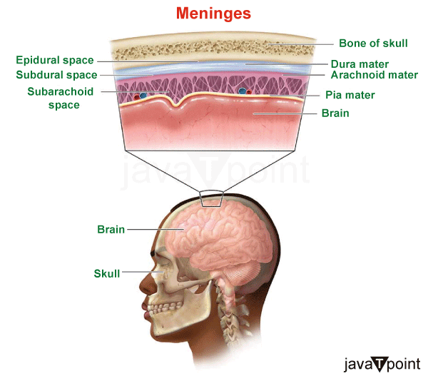
What is Meninges?Three membrane layers, meninges, surround and safeguard your central nervous system, including the brain and spinal cord. The dura mater, arachnoid mater, and pia mater membranes shield and support your brain as well as the blood arteries, nerves, lymphatics, and cerebrospinal fluid that envelops your central nervous system. In order to prevent the central nervous system (CNS) structures from coming into touch with the skull or spinal column, they wrap the CNS structures. By using anatomical position, the meninges may be found. The cranial meninges are the region that encircles the brain. The meninges of the spine, which cover the spinal cord and cauda equina. Types of Meninges1. Dura MaterThe periosteal layer, which is closest to the calvarium, and the meningeal layer, which is closest to the brain tissue, make up the dura, the outermost matter of the meninges. Together, these factors make the dura a thick, dense, fibrous membrane that is rather inelastic. The dura's strength comes from the periosteal layer, which is made up of fibroblasts and osteoblasts and contains a significant amount of extracellular collagen in the intercellular space. The only places where these two layers diverge are to create venous sinuses and dural reflections. When two face-to-face meningeal layers enter the cranial cavity to create the septa that divide the brain into compartments, this is referred to as a dural reflection. The falx cerebri and the tentorium cerebelli are the two primary dural reflections. The right and left cerebral hemispheres are divided by the falx cerebri, which has the form of a sickle and is linked to the roof of the skull. The tentorium cerebelli, which has a "U" form, is located between the cerebellum and occipital lobes. The tentorial incisura, also known as the tentorial notch, is an important aperture in the falx cerebri that permits the midbrain to pass through and into the middle cranial fossa. 2. Archanoid MaterThe cerebrospinal fluid (CSF) metabolism is carried out by the arachnoid, an avascular membrane that lies between the dura and the pia. In contrast to the pia mater, the arachnoid spans the cortical sulci rather than following their shapes. Its appearance varies depending on where in the skull it resides, but it is often a thin, translucent membrane. A surface mesothelial layer underneath the dura, a core layer of cells linked by several junction proteins, and a deep layer of less densely packed cells with numerous collagen fibers in their intercellular space make up its structure. According to evidence, arachnoid granulations, or groups of arachnoid villi, or parts of the arachnoid that protrude into the dura mater, may act as a network for communication between the systemic venous system and the CSF. 3. Pia MaterThe pia mater, the deepest layer of the meninges, is made up of two layers and, in contrast to the arachnoid, follows the shapes of the sulci and gyri. Collagen fibers are found in the pia's outermost layer, known as the epiphysial layer. In contrast, elastic and reticular fibers are found in the pia's innermost layer, known as the intima pia. The intima pia clings to the glial membrane, the outermost layer of neural tissue, while the mesothelial cells of the epiphysial layer link to the arachnoid mater through the arachnoid trabeculae. Notably, the cerebral pia mater creates sheaths around the blood arteries that go from the subarachnoid space to the brain parenchyma and enter and leave the brain perpendicular to the meninges. This sheathing generates an interstitial fluid-filled gap known as the perivascular or Virchow-Robin space between the vessel walls and the pia. Spaces within the Meninges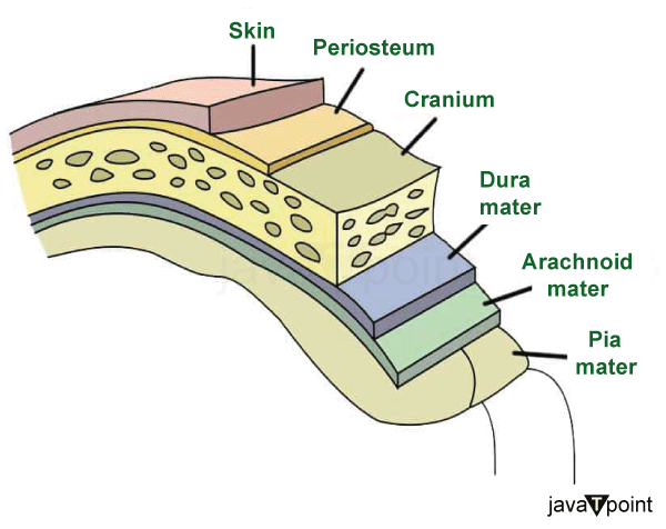
1. Epidural SpaceThe epidural gap may be located between the superior surface of the dura and the calvarium, which is the roof of the skull. The epidural space often occurs pathologically because the superficial layer of the dura, the periosteal layer, is tightly linked to the periosteum of the skull, despite the fact that arteries and veins flow between the inner surface of the calvarium and the dura. 2. Subdural SpaceThe area that could be present between the arachnoid mater and the meningeal layer of the dura is known as the subdural space. The subdural space, like the epidural space, is usually only present in pathological situations. 3. Subarchanoid SpaceThe area between the arachnoid and the pia, which is filled with cerebrospinal fluid, is known as the subarachnoid space. The choroid plexus in the brain's lateral ventricles generates CSF, which travels via the foramina of Luschka and into the subarachnoid space. CSF travels via the foramen magnum and down to the distal part of the spinal cord because the subarachnoid space connects the brain and spinal cord in a continuous fashion. The brain and spinal cord are protected from damage by the CSF, which also provides them with nourishment and removes waste. The brain's main arteries also pass through the subarachnoid region in addition to the CSF. Arachnoid trabeculae, which are strands of connective tissue from the arachnoid mater, also protrude into this area. Nerves Supply in MeningesThe vagus and trigeminal nerves are principally responsible for innervating the meninges, with specific cervical spinal nerves also playing a minor role. While a branch of the maxillary division of the trigeminal nerve supplies nerve supply to the dura of the middle cranial fossa, the anterior and posterior ethmoidal nerve branches of the ophthalmic division of the trigeminal nerve principally innervate the dura of the anterior cranial fossa. The tentorial nerve, which is also a branch of the ophthalmic division of the trigeminal nerve, principally innervates the tentorium cerebelli and the posterior portion of the falx cerebri. The infratentorial dura of the posterior cranial fossa is primarily innervated by the first three cervical spinal nerves and the cranial sympathetic trunk. Function of MeningesThe meninges serve as a barrier that protects the CNS's delicate organs from injury and serves as a support system for the CNS. The meninges :
The brain parenchyma may be temporarily and permanently harmed if these processes are disrupted. Clinical Significance of MeningesIn many pathological disorders, the meninges are involved. Bacteria penetrating the subarachnoid space, most often by the pathogens Streptococcus pneumoniae and Neisseria meningitides, may cause bacterial meningitis (BM), which results in inflammation of the meninges. Increases in intracranial pressure brought on by bacterial meningitis may cause neuronal damage; this is followed by parenchymal damage brought on by brain edema, hydrocephalus, and vascular changes. Additionally, research indicates that cognitive deficiencies may be caused by toxins generated by the microorganisms that have penetrated the body; this condition is treated with prompt antibiotic medication. The structure and function of the meninges are intimately tied to a joint group of disorders known as traumatic and atraumatic intracranial hemorrhages, which may be categorized according to where they occur within the meningeal layers. As an example, an epidural hematoma is a collection of blood in the restricted area between the dura and the skull (also known as the epidural space). This illness is often brought on by the rupture of the middle meningeal artery as a result of a temporal bone fracture brought on by head trauma. A CT scan reveals a lens-shaped biconvex hematoma as the hemorrhage quickly grows under arterial pressure and the dura separates from the calvarium. If left untreated, brain herniation caused by the hematoma's compression of the underlying brain tissue might result in death. Other intracranial hemorrhages include subdural hematomas, which often arise as a consequence of the rupturing of the bridging veins and the collection of blood in the possible subdural space between the dura, and arachnoid and subarachnoid hemorrhages, which take place in the subarachnoid space. Nontraumatic subarachnoid hemorrhages are sometimes further divided into traumatic and nontraumatic categories. Nontraumatic subarachnoid hemorrhages typically occur from an artery's spontaneous aneurysm (or rupture) in the space between the arachnoid and the pia. They are accompanied by an abrupt start of headache symptoms.
Next TopicParkinson Disease
|
 For Videos Join Our Youtube Channel: Join Now
Feedback
- Send your Feedback to [email protected]
Help Others, Please Share


