| |

Bacterial Skin InfectionThe major cause of skin diseases are bacteria, fungi, viruses, and parasites. The most common pathogen in bacteria is Staphylococcus aureus and beta-hemolytic Streptococci. Under viruses, Herpes simplex is proved to be the most common skin disease. Trichophyton rubrum has seen to be the prominent causative agent of skin and nail infections. 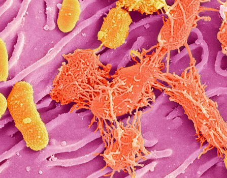
PathogenesisPrimary infections - Primary infections are featured by a single pathogen attack and occur typically on normal skin. These types of infections are caused by the effect of a single pathogen that leads to skin abnormalities. Primary infections can be easily detected as they have a visible area of infection and the required period of a disease. Some very common types of primary infections are Folliculitis, Impetigo, and Boils. Streptococcus aureus, Coryneform bacteria, and beta-hemolytic streptococci are the pathogens responsible for primary skin infection. The mode of entry of these bacteria is through the skin. Like if an insect bites you, the wound will become the entry site of these harmful pathogens. These infections include varicella, gonococcemia, measles, staphylococcal scalded skin syndrome. Even fungi of the special kind have a strong ability to invade tissue of keratin proteins and parts composed of keratin like nails, hair, and skin. Secondary infection - This infection occurs on the pre-existing area of infection on the skin. Due to the pre-existing infection, the clinical status and remedy period may vary according to the severity of the infection. The most common examples of secondary infection are toe web infection and Intertrigo. Clinical ManifestationsThe most common signs of skin infection include erythema and edema-like inflammation. Moreover, something severe can leads to the accumulation of furuncles (pus) or fluid in the infected site. A lesion can also be formed on the area without inflammation. Prevention and TreatmentThere are various methods to prevent/treat skin infections.
Methods for Laboratory DiagnosisCollection of specimen - There are different ways to collect specimen for bacteria, fungi, and viruses. For bacteriaSpecimens are collected from the infected area of the skin by a blade or by swabbing the skin. If there is a vesicle or pustule is present, the outermost layer is removed with an autoclaved surgical blade. Then, spread the pus or exudate on a clean glass slide and make a thin-smear-like layer for the Gram staining technique to identify the presence of gram-positive or gram negative bacteria. For ActinomycetesThe collection of specimen (pus) is done by the close lesion with autoclaved needle and syringe by the experienced staff. The exudates are collected from draining sinuses by holding the test tube right on the surface of the lesion which allows the pus and granules to flow seem lessly into the test tube. Granules are a form of aggregates of inflammatory cells, debris, protein substance, or filaments. Then, the pus and other exudates are observed under a microscope to identify the presence of granules in the material. For VirusesThe cleaning of vesicles are done using 70 percent ethanol which is followed by the use of sterile saline. The viral specimen is collected using the removal of the uppermost surface of the vesicle with a sterile needle or blade. Then, the cotton swab or tuberculin syringe is used to collect the fluids or exudates. The obtained fluid from vesicles contains a sufficient amount of viruses for culture. Direct smear of fluid can be made by scraping cells from the bottom of lesions. The fluid is smeared on a clean glass slide. Then fixation of cells is done by swinging the slide on the flame. The stains used for viruses are Giemsa or Wright stain or flourescein or peroxidase. For FungiThe samples made by cut wounds are obtained by scraping the infected skin into a sterile Petri dish or plate. Proper lesion of deep skin and subcutaneous tissue should be done by experiencing with a sterile blade and needle. For observation, mount the sample with two or three drops of physiologic saline or KOH and spread it on the glass slide. Then, cover the sample with glass coverslip ensuring no air spaces. CulturesThe bacteria that cause skin infections are cultured on artificial media. It is crucial to select the appropriate medium for specific bacteria. For general use, medium, if blood agar plates are suitable especially 5% defibrinated sheep blood. Many times selective medium are used along with general use medium for a better culture. In case of culturing of Staphylococcus aureus and Staphylococci pyogenes in a blood agar medium, S.aureus may overgrow and suppress the growth of S pyogenes when they are culture together. When S.pyogene and S.aureus is grown togther in a medium of blood agar and crystal violet, the S.pyogene overgrown S.aureus. Menungococci, gonocci, and brucellae culture must be incubated in atmosphere-rich CO2. If tuberculosis or fungus infection was found, the bacteria are collected on selective media and incubated aerobically. Primary InfectionsImpetigoThere are ways to identify impetigo on the basis of clinical histological and bacteriological investigation. The wound of common or superficial impetigo may have a group of beta-hemolytic streptococci, Staphylococcus aureus, or both. However, it is still not evident on which of these pathogens are responsible for the infection. The wound appear thick, adherent, dirty yellow surface, recurrent with an erythematous margin. Impetigo is very common in infants and highly contagious infection. It requires proper and quick treatment. Bullous impetigoOther form of impetigo is bullous impetigo caused by Staphylococcus aureus. The lesion of bullous impetigo are superficial thin walled and bullous. It can be distinguished from the common impetigo infection as it forms white, thin, transparent, varnish like surface appears on rupturing of lesion. This characterstics appearance on the lesion of bullous impetigo results from the action of epidermolytic toxin (exfoliation). The lesion are found in cluster in one area at a time. Ecthyma impetigoEcthyma is a form of impetigo which penetrate deeper into the skin. The scrape most often found on the legs and other parts of the body that are generally covered. They typically occur as serious infestation of pathogen. The ulcer have bulbous apperance when the outer surface and pus are removed. This infection cause lesion that heal slowly and leave scars behind. Cellulitis and ErysipelasCellulitis are a diffuse inflammation of loose connective tissue mainly subcutaneous tissue and is affected by Streptococcus pyogenes. The invasion of S pyogene is generally through a breach in the surface of skin. The infection is facilitated by the presence of edema. Cellulitis can occur in normal skin. However, the lesion are big, tender, poorly defined margins. It is difficult to differentiate between Streptococcal cellulitis and Straptococcal erysipelas. When clinally tested, it is found that erysipelas is more trivial, as the lesion has sharp margin compared to the poorly defined margins of cellulitis. The infection usually occur on the cheeks. Staphylococcal Scalded Skin SyndromeStaphylococcal scalded skin syndrome is also referred to as Lyell's disease or epidermal necrolysis. The infection begins as localised lesion which is followed by large erythema and exfoliation of the skin. This disease is caused by phage type 2 staphylococci which widespread the epidermolytic toxin. It is more usual in infants than adults. FolliculitisThis can be classified into two broad groups based on their location i.e. deep and superficial. The most superficial form of folliculitis is Staphylococcus folliculitis, manifested by minute erythematous follicular pustules without involving the surrounding area. The favorite site of the disease to occur is scalp. The gram negative folliculitis occurs majorly as super infection in patients with acne vulgaris. These patients require long term, systemic antibiotic therapy. These pustules are grouped around the nasal area. The causative agent in found around nostrils and pustules. The Propionibacterium acnes folliculitis has been misunderstood by S.folliculitis. The primary lesion is of white to yellow follicular pustules, flat or curved. Gram stain procedure indicates several intracellular and extracellular gram-positive pleomorphic rods. These lesion occur more commonly in men than women. The process initate at the age of adolescence, when acne appears yet inmany cases it occurs years later. In deep folliculitis,the infection extends deeply into follicle, and it results in a more distinct inflammatory response than that visible in superficial folliculitis. In sycosis barbae, hair peirce the primary lesion follicular pustules. Beard men may be more prone to this infection than clean shaved men. A boil (furuncle) is an infection of follicle caused by Staphylococcus. It involved subcutaneous tissues. The most occuring sites of boil are the hairy parts of areas that are opened to friction and maceration. A carbuncle is a confluence of boil, a large painful lesion with multiple sites from where exudates will drain. Erysipeloidit is a infection that it usually e occurs in in workers in fishery industry fisherman and meat Handler. it causes redness of skin mainly the fingers or the back of the hand. It is a kind of bacterial infection which stays up to several days to weeks. Erysipeloid is caused by Erysipelothrix rhusiopathie. Pitted keratolysisIt is an infection that occurs on surface of skin producing a protrusion like pits. The pits may merge into irregularly structured areas of superficial erosion. These pits are formed by lytic cycle of bacteria and mainly spreads on borders of the infected area. The main areas that get caught by the infection of bacteria are heels, ankle, the volar pads, and toes. The chance of getting infected is enhanced by the aggravating factors like humidity and extreme temperatures. Gram-positive Coryneform bacteria are the main causative agent of Pitted keratolysis. ErythrasmaErythrasma is a severe, superficial infection that occurs in the area like the toe web, groin, axilla, pubis, and mammary folds. Most contusions are asymptomatic however some are mildly symptomatic with inflammatory and itching effects. The infected area appears as an irregular patchy surface, dry and scaly. These patches are pink in the beginning and turn brown. The spread of the kind of infection is more common in hotter climates. The main causative agent is Corynevacterium minutissimum. As the bacteria are small in size, which makes it even more difficult to observe in the KOH sample of infected scales. However, it is observable with gram staining technique. The common morphological feature is coral red fluorescence under wood's light. TrichomycosisTrichomycosis comprises the hair in the axillary and pubic areas and is characterized y the establishment of nodules of different consistency and color. The disease is generally asymptomatic and not contagious. The skin beneath the region is normal. The infected hairs are collected and placed on a slide for microscopic observation with a drop of 10% KOH under a coverslip. The nodules on the hairs are made of short bacillary forms. Trichomycosis re-associated with three types of corneyforms, i.e. C.minutissimum, lipolytic and C.tenuis. Secondary InfectionsIntertrigoIt is a type of secondary infection that commonly occurs in chubby infants or adults with obesity. Obese people have thick skin that folds and generate heat and moisture on rubbing which produces erythema , maceration and erosions. This can also be produced by the overgrowth of flora. Acute Infectious Eczematoid DermatitisAcute infectious eczematoid dermatitis arises from a primary scrape like a boil or an ear or nose that drain which is a source of infectious exudates. A diagnostic feature of this disease is a streak of dermatitis along the border of the flow of the discharge exudates. The most frequently isolated organisms for this disease is staphylococcus. Pseudofolliculitis of the BeardPseudofolliculitis of the beard is a common disease that occurs mostly in the area of the beard in people who shave rarely. The characteristic scrape is typically erythematous papules or rarely pustules obtaining buried hairs. This disease arises when strongly curved hair emerges from curved hair follicles and reenters the skin to produce ingrown hair. The pathogen which commonly seen to cause this disease is gram-positive microorganisms that belongs to resident flora. A clear idea for the non-pathogenic bacteria attack when the host defense is not active. Toe Web InfectionToe web infection or disease is commonly known as athlete's foot. This disease was traditionally referred to as fungal infection. This assumption has been taken into account again because fungi cannot recover from the lesion throughout the course of the disease. Scientists have started to believe that the dermatophytes, the first invaders are responsible I cause skin damage that allows bacterial overgrowth which enhances the infection of hyperkeratosis and maceration.
Next TopicBacterial diseases in plant
|
 For Videos Join Our Youtube Channel: Join Now
Feedback
- Send your Feedback to [email protected]
Help Others, Please Share


