| |

Blood Supply to the BrainAround 2% of the body's weight is made up of the brain, which still gets 15% to 20% of the heart's entire output. The brain is mostly dependent on oxidative metabolism and has a rather high metabolic requirement. Ten seconds after the arterial blood supply to the brain is cut off, a person loses consciousness, and just a few minutes later, irreversible brain damage has already been done. The anterior and posterior circulations of the brain's arterial blood supply may be distinguished. Both the left and right vertebral arteries and the left and right internal carotid arteries are the sources of the first. Like all other bodily systems, the central nervous system has to be continuously nourished and oxygenated. At rest, the brain consumes one-fifth of the body's total oxygen intake, indicating a particularly high oxygen requirement for the organ. As a consequence, ischemic cell death occurs within minutes of oxygen deprivation, which is also particularly sensitive to this tissue. Cerebral Circulation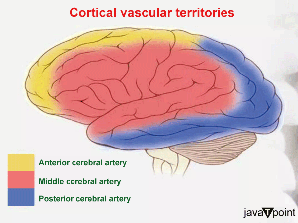
The flow of blood through the brain's vascular system, or cerebral circulation, is what keeps the brain supplied with blood. In an adult human, cerebral blood flow is generally 750 milliliters per minute or 15% of cardiac output. The blood vessels supply the brain with oxygenated blood, glucose, and other nutrients. In order to eliminate carbon dioxide, lactic acid, and other metabolic waste products, veins return "used or spent" blood to the heart. The neurovascular unit controls cerebral blood flow so that activated neurons may get the proper quantity of energy at the appropriate moment. The cerebral circulatory system contains precautions, such as autoregulation of the blood arteries, since the brain would soon suffer damage from any cessation of blood flow. A stroke might happen if these protections are ineffective. The cerebral blood flow is the measure of blood flow in the brain. The perception of gravity forces by bodies is altered by sudden, powerful accelerations, which may also seriously impede cerebral blood flow and other regular bodily processes to the point of posing a major risk to one's life. The following explanation is based on a hypothetical depiction of human cerebral circulation. Different creatures have different circulatory systems with different nomenclatures. According to the several arteries that feed the brain, the blood flow to the brain is often split into anterior and posterior parts. The vertebral and internal carotid arteries are the two major arteries that feed the anterior and posterior brains, respectively. The anterior and posterior cerebral circulations are joined through the bilateral posterior connecting arteries. They are a component of the Circle of Willis, which gives the brain backup circulation. The Circle of Willis establishes links between the anterior and posterior cerebral circulation along the floor of the cerebral vault, delivering blood to tissues that would otherwise become ischemic in the event that one of the supply arteries is blocked. Anterior Cerebral CirculationAll the arteries that emerge from the internal carotid arteries are part of the anterior circulation. It is in charge of the blood flow to the front and center of the brain. The following arteries make up the anterior circuit:
1. Internal Carotid ArteryA branch of the common carotid artery, the internal carotid artery, is one of two. The anterior and middle regions of the brain get a significant amount of their nutrition from it. The internal carotid artery is now divided into four sections: the cervical portion in the neck, the petrous portion at the base of the skull, the cavernous portion within the cavernous sinus, and the intracranial portion above the cavernous sinus. The following distinctions may be seen between the Cincinnati categorization and the new system:
2. Anterior Cerebral Artery Comparatively speaking to the middle cerebral artery, the anterior cerebral artery (ACA) is a much smaller branch of the internal carotid artery. The medial region of the Sylvian fissure is where it starts, near the end of the internal carotid artery, after the ophthalmic branch leaves the body. On its way to the longitudinal cerebral fissure, it follows an anteromedial path, moving ahead of the optic nerve (CN II). By way of the brief anterior communicating artery (AComm), it anastomoses with the oblique counterpart here. The paired arteries then pass through the longitudinal cerebral fissure that runs along the corpus callosum's genu. 3. Anterior Communicating ArteryA small, narrow blood vessel called the anterior communicating artery (AComm) connects the anterior cerebral arteries horizontally. The vessel is situated anterior to the optic chiasm and posteromedial to the olfactory tracts, crossing the ventral side of the median longitudinal fissure. The anterior bridge connecting the left and right sides of the anterior circuit is formed by this vessel. Additionally, it completes the Willis circle, the anterior portion of the anastomotic ring. 4. Middle Cerebral ArteryThe greatest terminal branch of the internal carotid artery is called the middle cerebral artery (MCA). On the Reil (insula) island, it passes through the Sylvian (lateral) fissure before coursing in a posterosuperior direction. It then separates to provide the lateral cortical surfaces and the insula. The brain's central and cortical areas get multiple tributaries from the vessel. The lenticulostriate arteries, which feed the lentiform nucleus and the posterior limb of the internal capsule, are among the relatively tiny central branches that pass through the anterior perforated material. 
Posterior Cerebral CirculationAll blood veins that emerge from the vertebrobasilar system are referred to as being in the posterior circulation. These blood veins supply the occipital lobe of the brain and the hindbrain. These are some of the vessels in the posterior circuit:
Vertebral ArteryThe foramen magnum, which is located anteromedial to the brainstem, allows the vertebral arteries access to the cranial vault. Each vertebral artery's branches relate to:
Basilar ArteryAn essential vessel located in the pontine cistern is the basilar artery. As it ascends in the basilar groove, it is anterior to the pons and posterior to the clivus. The pons, cerebellum, internal ear, and other surrounding structures are supplied by its branches. The basilar artery has three main branches:
The artery's lateral surface and distal bifurcation, respectively, give birth to the smaller pontine and posteromedial (paramedian) arteries. The basilar artery splits into two posterior cerebral arteries at its terminus. The union of these vessels posteriorly completes the circle of Willis with the posterior connecting arteries. Posterior Cerebral ArteryThe basilar artery splits into two terminal branches, including posterior cerebral arteries (PCA). Behind the dorsum sellae is where the division is located. The oculomotor nerve (CN III) separates it from the superior cerebellar artery. The artery keeps moving in a direction that is lateral to the midbrain (near the trochlear nerve, CN IV). The posterior communicating artery that it emits completes the Willis circle. The vessel then keeps moving towards the tentorial aspect of the cerebrum while navigating the cerebral peduncles. The occipital and temporal lobes are supplied here. Posterior Communicating ArteryA long, narrow arterial that emerges from the posterior cerebral artery is known as the posterior communicating artery (PComm). It is substantially longer than the anterior communicating artery, which is its anterior counterpart. The vessel lies lateral to the mammillary bodies of the hypothalamus and medial to the uncus of the temporal lobe. The optic tract's proximal portion and the vessel's distal portion may overlap. The Willis circle is completed posteriorly by the posterior connecting artery. Additionally, it provides tributaries to the thalamus, internal capsule, cerebral peduncles, and optic tract.
Next TopicBrain Cancer Symptoms
|
 For Videos Join Our Youtube Channel: Join Now
Feedback
- Send your Feedback to [email protected]
Help Others, Please Share


