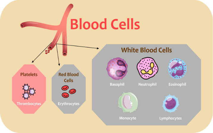
Blood CellsHuman blood cells are produced during cell differentiation and are found in connective tissue, known as blood. The bone marrow produces blood stem cells, which are the precursors to all human blood cells. Blood accounts for 45 % of the average tissue volume; the remaining 55% comprises plasma and liquid blood. The three main types of blood cells are:

1. Erythrocytes (Red Blood Cells)Red blood cells are the most popular cell type in the human body. They make up approximately 40 to 45% of the blood. They have a relatively thin bowl-shaped small, rounded disc-like structure with a diameter of 6.2-8.2 micrometres. They have a thick rim around them and a narrow, submerged centre. The nucleus is absent in RBCs. They can alter shape without breaking. Erythropoietin helps to control RBC production. RBCs contain 33 % of the total amount of haemoglobin. The iron in haemoglobin is responsible for the red colour of blood. RBCs cannot repair themselves. A Red Blood Cell has a life cycle of 120 days. An adult generates 2.4 million new erythrocytes per second. In males, the quantity of RBCs varies from 4.3-5.9 million per mm³, whereas, in females, the amount of RBCs varies from 3.5-5.5 million per mm³. Functions: The two main functions of RBC are to carry oxygen from the lungs to other parts of the body, take up carbon dioxide from the tissues, and leave it in the lungs. 2. Leukocytes (White Blood Cells)Leukocytes make up about 1% of the blood. The number of leukocytes count is 4500-11,000 m3. They make up most of the immune system. It is the part of the body that defends itself against foreign pathogens and infections. They are developed in the bone marrow by multipotent cells called hematopoietic stem cells. They can be found throughout the body, including connective tissue, lymphatic vessels, and the bloodstream. Leukopenia is a low white blood cell count induced by bone marrow damage done by medicines, radio waves, or chemotherapy. Leukocytosis is a high white blood cell count triggered by various conditions, such as infectious diseases and autoimmune disorders in the body. WBCs are of two types- Granulocytes and Agranulocytes. Granulocytes are cells with visible granules or grains inside, and Agranulocytes are devoid of visible grains under the microscope. Granulocytes are further subdivided into Neutrophils, Eosinophils and Basophils. Agranulocytes are subdivided into Lymphocytes and Monocytes. A. Neutrophils They are the most predominant type of white blood cell. They have a nucleus with multiple lobes. The cytoplasm consists of fine granules. The neutrophil count ranges from 2000 to 7500 cells per mm3. They are also known as polymorphonuclear due to their variety of nuclear shapes. The diameter ranges from 10 to 12 m. The average lifespan varies between 6 hours and a few days. Functions: Neutrophils use phagocytosis to kill bacteria. They also produce superoxides which can kill a wide range of pathogens. B. Eosinophils The count of Eosinophils is 40-400 cells per mm³. They have large granules. The nucleus is divided into two lobes. The diameter of a cell is 10 to12 μm. The life cycle of these cells is 8 to 12 days. Functions: They kill parasites and have a role in allergic reactions. They release toxins from their granules to kill pathogens. C. Basophils Basophils count varies from 0-100 per mm3. They look Colourful when stained and visualised under the microscope. They have a pale nucleus that is usually hidden by granules. Bi-lobed or Tri-lobed nucleus present. The diameter of these cells is 12-15 μm. Accounts for 0.4%. Life span varies from hours to a few days. Functions: It is used in allergic reactions. Produce anticoagulants and antibodies that protect the bloodstream from hypersensitivity. Basophils contain histamine, which causes blood vessels to dilate, allowing more immune cells to reach the injury site. Secrete heparin, an anticoagulant that seeks to promote other WBC mobility by trying to prevent clotting. D. Lymphocytes They are small oval cells, and the nucleus is present. The count is 1300 to 4000 per mm3. The diameter of a cell is 7-8 μm to 12-15 μm. Accounts for 30% of the total cells. The life cycle is of years for memory cells and weeks for other varieties. Functions: T lymphocytes are responsible for cell-mediated immunity. B lymphocytes are in charge of host defence or antibody production. They can recognise and memorise infecting bacteria and viruses. Perform the task of destroying cancer cells. They present antigens to activate other cells of the immune system. E. Monocytes The white blood cell with a giant nucleus, which is kidney-shaped. The count ranges between 200 and 800 monocytes per millilitre. They become macrophages after escaping the bloodstream. The diameter ranges from 15 to 30 micrometres. Monocytes makes up 5.3% of total blood cells. The life cycle of these cells ranges from hours to a few days. Function: Enters the tissue, where they grow larger and become macrophages. Cleanse the body of old, destroyed, and dead cells. 3. PlateletsA nucleus in the Platelet is missing. These are small fragments of bone marrow. 150,000-400,000 platelets are present in each microliter of human blood. Functions: Platelets are clotting cell fragments produced by the body. Promotes other blood clotting mechanisms. Secrete procoagulants to improve blood clotting. They secrete vasoconstrictors, which constrict blood vessels, causing vascular muscle spasms in damaged blood vessels. They secrete chemicals that attract neutrophils and monocytes to the inflammation site. Dissolve blood clots when they are no longer required. Bacteria are destroyed and digested. Helps in the digestion and destruction of bacteria. They secrete growth factors to keep the inner lining of blood vessels healthy.
Next Topic#
|
 For Videos Join Our Youtube Channel: Join Now
Feedback
- Send your Feedback to [email protected]
Help Others, Please Share


