| |

Brain Parts and FunctionsOverviewA miraculous three-pound organ, the brain directs all bodily processes, deciphers data from the outside world, and embodies the spirit and mind. The brain controls a wide range of processes, including intelligence, creativity, emotion, and memory. The cerebellum, brainstem, and cerebrum comprise the brain, shielded within the skull. Our five senses-sight, smell, touch, taste, and hearing-receive data from the brain, sometimes many at once. It puts the messages together in a manner that makes sense to us and can be remembered. Thoughts, speech, memory, arm and leg movement, as well as the operation of several bodily organs, are all controlled by the brain. The spine and brain together make up the central nervous system (CNS). Spinal nerves, which branch off the spinal cord, and cranial nerves, which branch off the brain, make up the peripheral nervous system (PNS). What is Brain?The brain is a sophisticated organ that manages all bodily functions that keep us alive, including cognition, memory, emotion, touch, motor skills, vision, respiration, temperature, and hunger. The central nervous system, or CNS, is comprised of the brain and the spinal cord that projects from it. 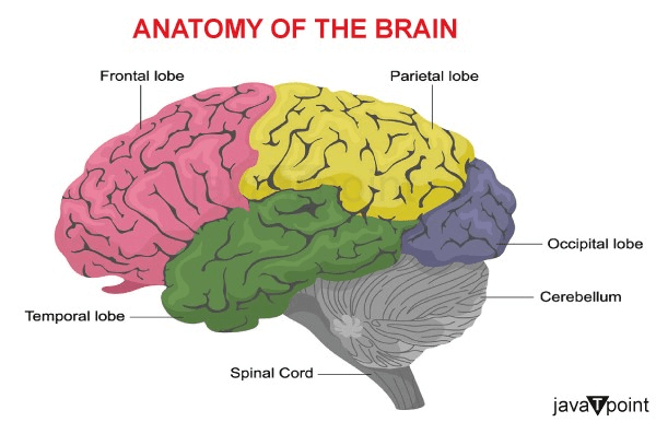
The typical adult's brain weighs 1 to 1.5 kg and contains roughly 60% fat. Water, protein, carbs, and salts make up the remaining 40% of the body. The brain does not function like a muscle. It includes neurons, glial cells, blood vessels, and nerves. According to recent estimates, the brain has between 86 billion and 100 billion neurons. The central nervous system is made up of the spinal cord and the brain. It is in charge of thinking, interpretation, and where the control for bodily motions comes from. The skull, which protects the brain frontally, laterally, and dorsally, encloses it. There are 22 bones in the skull, of which 14 make up the face bones and the other 8 the cranial bones. The cerebrospinal fluid surrounds the brain, which is anatomically enclosed within the skull. Hollow areas on the surface of the brain are filled with a fluid called cerebrospinal fluid (CSF), which circulates within the skull and spinal cord. The specialist ependymal cells create 500 mL or less of cerebrospinal fluid each day. The main job of the CSF is to protect the brain by absorbing mechanical shocks and taming little jolts. Additionally, it gives the brain fundamental immunological defense. Furthermore, CSF gives the brain buoyancy. Therefore, the weight of the brain is essentially nullified since it is floating in a layer of CSF. The weight of the brain would prevent it from moving freely if it weren't floating in CSF, which would shut off blood flow to the bottom half of the brain. Neurons in the impacted region would die as a result. Brain coverings: MeningesThe meninges, which are made up of three layers, surround the spinal cord and brain.
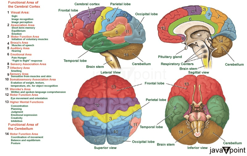
Parts of the BrainThe brain structure is composed of three main parts: the forebrain, midbrain, and hindbrain, each with multiple parts. 1) ForebrainIt is the largest part of the brain. It is furthermore divided into three parts: a. Cerebrum The greatest portion of the human brain, the cerebrum, sometimes referred to as the cerebral cortex, is connected to higher-order mental processes, including cognition and action. The grey surface, which is a bit thicker than our thumb, is composed of nerve cells. Under the skin, white nerve fibers transmit impulses between nerve cells in different regions of the brain and body. The neocortex, a mammal-specific six-layered structure, has a wrinkled surface that enhances its surface area. It has four "lobes" that make up each portion. The four of them are the frontal lobe, parietal lobe, occipital lobe, and temporal lobe. Types and Functions of the Lobes:
Two parts, or hemispheres, make up the cerebral cortex. Gyri and folds (sulci) are seen all over it. The interhemispheric fissure, also known as the medial longitudinal fissure, goes from the front of the head to the rear of the body and is where the two parts come together. The left hemisphere governs the right side of the body, while the right hemisphere controls the left. The corpus callosum is a large, C-shaped structure of white matter and nerve channels that connects the two halves. In the middle of the cerebrum lies the corpus callosum. Grey matter and white matter are the two different tissue types that make up the brain. The majority of the brain's grey matter is made up of several cell types. Axons, which link the different grey matter regions of the brain together, make up the majority of white matter. The cortex or cerebral mantle refers to the outside of the brain. The cortex has a huge surface area as a result of its high convolution. In the cerebrum, there are also:
b. Thalamus The thalamus, a tiny structure just above the brain stem, transmits sensory information from the sense organs. Additionally, it is in charge of communicating coordination and movement-related motor data. The limbic system of the cerebrum is where the thalamus is located. The fundamental functions of this limbic system are creating new memories and storing prior events. c. Hypothalamus Exactly underneath the thalamus in the brain, there is a tiny but crucial region known as the hypothalamus. It is regarded as the brain's major area since it performs the following tasks:
2) MidbrainNear the center of the brain, the midbrain is situated above the hindbrain and under the cerebral cortex. It consists of many nuclei and fasciculi, as well as the tectum, tegmentum, cerebral aqueduct, and cerebral peduncles. The midbrain's main function is to serve as a relay station for our auditory and visual systems. There are many dopamine-producing neurons in the midbrain regions known as the substantia nigra and red nucleus, which are also important in controlling bodily movement. Parkinson's disease is correlated with neuronal degeneration in the substantia nigra. The smallest part of the brain, the midbrain, is situated in the middle of the cranial cavity. Midbrain has two main parts:
3) HindbrainOne of the three primary brain areas is the hindbrain, which is situated in the bottom back of the brain and develops from the rhombencephalon. It contains the majority of the brainstem as well as the cerebellum, a thick structure resembling a coral reef. Because it links the brain to the spinal cord and controls several essential processes, including respiration and heartbeat, the brainstem is one of the most crucial components of the complete central nervous system. The 12 cranial nerves are mostly located in the hindbrain. The pons, cerebellum, and medulla oblongata are the primary structures of the hindbrain. a. Cerebellum The cerebellum, which is housed in the medulla and pons' posterior region, is the second-largest component of the brain. The cerebellar tentorium and transverse fissure divide the cerebrum from the cerebellum. The folia are parallel ridges that run along the cortex, which is the cerebellum's outer surface. The cerebellum also consists of cerebellar peduncles, nuclei, and anterior and posterior lobes. The outer grey cortex and the inner white medulla are the two hemispheres that make up the cerebellum. During walking, running, cycling, swimming, and finely controlling voluntary motions, it coordinates and maintains the body's equilibrium. The cerebellum's primary tasks are as follows:
b. Pons The pons, which link the remainder of the brainstem to the cerebral cortex, receives its name from the Latin word for "bridge." It is bulbous in form and located directly underneath the midbrain. It functions as a hub for the coordination of impulses and communications that go between the two hemispheres of the brain and the spinal cord. The pons is home to four cranial nerves: the abducens nerve, which aids in eye coordination; the facial nerve, which coordinates movement and sensation in the face; the vestibulocochlear nerve, which processes sounds and aids in balance; and the trigeminal nerve, which coordinates chewing and carries sensory data from the face and head. c. Medulla Oblongata The medulla oblongata is the bottom portion of both the brainstem and the whole hindbrain, and it's where the brain connects to the spinal cord. Although the medulla is just 3 cm long, it is a critical nerve tract that houses the control centers for our autonomic vital processes, such as heart rate, blood pressure, breathing, and numerous involuntary responses like swallowing and sneezing. White and grey matter coexist in the medulla, which is also the location of four cranial nerves: the glossopharyngeal nerve, which coordinates some taste sensations and mouth movements; the vagus nerve, which regulates voice, gag reflex, and mouth movements; the accessory nerve, which regulates head and neck movements; and the hypoglossal nerve, which regulates tongue movements and muscles used in speech. Deeper Structures within the Brain
The pituitary gland, a pea-sized organ located deep inside the brain beneath the nose's bridge, is often called the "master gland." By controlling the release of hormones from the thyroid, adrenals, ovaries, and testicles, the pituitary gland controls the operation of other glands in the body. It gets chemical cues from the hypothalamus through its blood supply and stalk.
There is an amygdala beneath each hemisphere of the brain, which are tiny, almond-shaped structures. The limbic system's amygdalae, which also controls emotion and memory and are linked to the brain's reward system, stress, and the "fight or flight" reaction when someone detects a danger, are part of the limbic system.
The hippocampal formation is a bigger structure that includes the hippocampus, a curving seahorse-shaped organ on the bottom of each temporal lobe. It promotes spatial awareness, memory, learning, and navigation. It gets information from the cerebral cortex and might be involved in the development of Alzheimer's disease.
A stalk connecting the pineal gland to the third ventricle's top is found deep within the brain. The pineal gland produces melatonin in response to light and dark, which controls circadian rhythms and the sleep-wake cycle.
Four open spaces connected by tunnels exist deep inside the brain. The space below the meninges' arachnoid layer and the central spinal canal are likewise accessible via them. Cerebrospinal fluid, or CSF, is produced by the ventricles and flows between the meninges as well as in and around the ventricles, the spinal cord, and both. The spinal cord and brain are encircled by CSF, which also serves to transport nutrition and clean away waste and pollutants. Blood Supply in the BrainThe vertebral and carotid arteries are the two groups of blood vessels that feed the brain with blood and oxygen. You may feel your pulse when you press your fingers on the place where the external carotid arteries run up the sides of your neck. The internal carotid arteries branch into the skull and supply the brain's frontal lobe with blood. The basilar artery, which carries blood to the back of the brain, is made up of the vertebral arteries, which merge at the brainstem after traveling through the spinal column into the skull. Blood travels from the front of the brain to the back, and the circle of Willis, a loop of blood vessels towards the bottom of the brain that links major arteries, facilitates communication between the arterial systems. Cranial NervesThere are 12 cranial nerves, often known as the dome of the skull, within the cranium:
The first two cranial nerves emerge from the cerebrum, while the next 10 nerves come from the brainstem, which comprises the midbrain, pons, and medulla. Cells of the BrainNeurons and glial cells are the two different kinds of cells that make up the brain. Nerve cellsNeurons come in various sizes and forms, but they always have a cell body, dendrites, and an axon. The neuron sends information through chemical and electrical impulses. Think about the wiring in your house. A light bulb will shine when a light switch is switched on thanks to the multiple wires that make up an electrical circuit. When stimulated, a neuron will transfer its energy to nearby neurons. A synapse is a very small opening via which neurons may communicate with one another or "talk" (Fig. 12). Dendrites, the several arms that make up a neuron, are like antennae that a neuron uses to take up signals from neighboring nerve cells. These signals are sent to the cell body, which decides whether or not to send them on. Important signals are sent to the end of the axon, where neurotransmitter-filled sacs emerge to form the synapse. The neurotransmitter molecules enter the receiving nerve cell via the synapse and bind to certain receptors, stimulating the cell to deliver the message. Glia CellsThe brain's glial cells, named after the Greek word for glue, feed, shield, and support neurons structurally. Glia is the most prevalent kind of cell in brain tumors and has between 10 and 50 times more of them than nerve cells.
Why is Brain Important?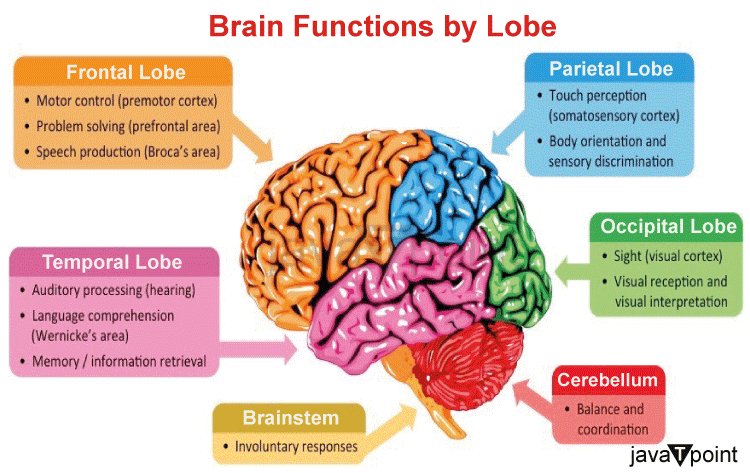
The brain serves as the nervous system's command center. A person's personality, mobility, eyesight, sleep, and other critical biological processes may alter when distinct areas of the brain are damaged. For example, brain death may result from a stroke or severe brain damage. Although they will never recover awareness, a person who has experienced brain death is nonetheless technically alive. The patient is put on artificial life support in order to maintain life by keeping the heart and lungs functioning. The heart continues to beat for a brief period of time after brain activity ends because it has a distinct electrical system from the brain. Before diagnosing brain death, physicians will do a number of tests. Everyone concerned suffers from the trauma of brain death. It may be difficult to accept the prognosis when a loved one is still breathing and exhibiting signs of life. The cerebellum, brain stem, and cerebrum make up the three primary parts of the brain. The brain controls several biological processes by distributing chemical and electrical impulses throughout the body, and it also detects changes in the environment. Most of the body's functions are communicated through the spinal cord on behalf of the brain. Billions of nerve cells across the CNS are used to do this.
Next TopicBrain Tumour Causes
|
 For Videos Join Our Youtube Channel: Join Now
Feedback
- Send your Feedback to [email protected]
Help Others, Please Share


