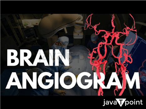
Brain AngiogramA type of angiography called cerebral angiography offers images of the blood arteries in and around the brain, making it possible to find anomalies such aneurysms and arteriovenous malformations. The University of Lisbon's Egas Moniz, a Portuguese neurologist who also contributed to the creation of thorotrast for use in the treatment, invented it in 1927. The carotid artery is often reached by inserting a catheter into a major artery (such the femoral artery), which is then threaded through the circulatory system to provide a contrast agent. A first series of radiographs is obtained as the contrast material travels through the arteries of the brain, and a second series is taken as it enters the veins. 
Compared to less invasive techniques like computed tomography angiography and magnetic resonance angiography, cerebral angiography may produce better images for some purposes. Additionally, based on its results, cerebral angiography enables some treatments to be started right away. Due to the development of endovascular therapeutic procedures, cerebral angiography has recently taken on a therapeutic significance. Embolisation, a less invasive surgical method, has grown in significance in the multimodal treatment of cerebral MAVs since it facilitates further microsurgical or radiosurgical therapy. Another kind of treatment made available by angiography (if the images reveal an aneurysm) is the insertion of metal coils through the catheter already in situ and manoeuvred to the aneurysm site. These coils promote the development of connective tissue at the area over time, reinforcing the vessel walls. In some legal systems, a cerebral angiography is necessary to prove brain death. Prior to the development of contemporary neuroimaging techniques like MRI and CT in the mid-1970s, cerebral angiographies were frequently used as a tool to infer the existence and location of specific types of lesions and hematomas by looking for secondary vascular displacement caused by the mass effect associated with these medical conditions. A variety of main cerebral disorders can now be directly seen using sophisticated non-invasive diagnostic techniques, rendering the use of angiography as an indirect assessment tool obsolete. However, it is still often used to assess different vascular diseases in the skull. UsesThe diagnosis made by cerebral angiography may be followed in the same location by therapeutic treatments. Different intracranial (inside the skull) or extracranial (outside the head) illnesses are imaged with cerebral angiography. Examples of intracranial diseases include non-traumatic subarachnoid haemorrhage, non-traumatic intracerebral haemorrhage, intracranial aneurysm, stroke, cerebral vasospasm, cerebral arteriovenous malformation (for Seltzer-Martin grading and plan for intervention), dural arteriovenous fistula, embolisation of brain tumours like meningiomas, and cavernous sinus haemorrhage. Carotid artery rupture, carotid artery stenosis, cervical spine trauma, nasal bleeding, subclavian steal syndrome, and the intention to embolise adolescent nasopharyngeal angiofibroma before surgery are examples of extracranial illnesses. Although cerebral angiography, which has been used widely in the evaluation of intracranial sickness, provides a higher resolution on the conditions of blood vessel lumens and vasculature than Computed Tomography Angiography (CTA) and Magnetic Resonance Angiography (MRA). The gold standard for finding intracranial aneurysms and determining whether endovascular coiling is feasible is cerebral angiography. It is possible to perform a cerebral angiography through the radial or femoral arteries to treat cerebral aneurysms with a variety of devices. This technique is not recommended for people with certain health issues, including contrast allergy, renal insufficiency, and coagulation abnormalities. Technique Prior to the procedure, a thorough neurological and history assessment is carried out, together with a review of the blood parameters and any accessible imaging. Arch anatomy and variances are considered when analysing imaging in order to choose the best catheters to examine the veins. To confirm that the subject has an appropriate level of haemoglobin and to rule out sepsis, the whole blood count is examined. When assessing serum creatinine, renal impairment is ruled out. Prothrombin time is measured in the interim to rule out coagulopathy. An informed consent is given after being informed of the procedure's hazards. Anticoagulants are avoided wherever possible. For diabetics who are fasting, the amount of insulin required is cut in half six hours before the surgery. The left arm/forearm and bilateral groynes are prepped for femoral artery access and brachial artery/radial artery access, respectively. Prior to sedation or anaesthesia, the patient's neurological condition is noted. If the person is agitated or in pain, sedatives such intravenous midazolam and analgesics like fentanyl may be administered. After that, the individual is placed in a supine position with their arms by their sides. Subjects who are uncooperative may have their foreheads touched to stop movements. It is advised that the subject maintain complete stillness, especially when fluoroscopy images are being taken. Additionally, it is important that the subject not swallow while having their neck photographed. To lessen motion artefact in the photos, several steps are taken. The recommended site of entry is through the Right Femoral Artery (RFA). Brachial artery access is chosen if RFA access is not ideal. It is possible to utilise an 18G access needle or a micropuncture system with or without ultrasound guidance. There are four different kinds of catheters that can be used: an angled vertebral catheter for routine instances, a Judkins right coronary catheter (Terumo) for vessels with twists and turns, a Simmons catheter and a Mani's headhunter catheter (Terumo) for severely twisty vessels. To avoid clotting around the 5Fr sheath, the area is also lined with heparinized saline and flushed. Terumo hydrophilic Glidewire 0.035 inches can be used as a guidewire. "Double flush" and "Wet Connect" techniques are employed to prevent embolism (caused by a blood clot or an air embolism). The "double flush" procedure involves aspirating blood from the catheter using a saline syringe. The catheter is then flushed with a second syringe of heparinized saline. The process known as "wet connect" joins a syringe to a sheath without air bubbles. The primary method for visualising the cerebral blood arteries is digital subtraction angiography. Advance the catheter over the guidewire. Additionally useful is rotating the catheter while advancing it. Using a "roadmap" to advance catheters or guidewires before any vessel bifurcation can help to prevent vessel dissection by superimposing a previous image on a live fluoroscopic image. Although cerebral angiography offers a higher resolution on the. Once the catheter is in place, the guidewire is progressively removed while heparinized saline drips into the catheter simultaneously to prevent air embolism. To prevent wedging, dissection, or intracatheter clotting, the catheter's backflow should be established prior to contrast administration. Extra caution should be used when catheterizing the vertebral artery to avoid vascular dissection or vasospasm. Vasospasm or dissection could be indicated by a delayed or partial contrast washout. View RadiographicA cervical arch angiography is carried out if there is any indication of aortic arch narrowing or any structural changes, such as bovine arch (the brachiocephalic trunk comes from the same location as the left common carotid artery). The primary branches of the aortic arch are difficult to cannulate if such an anomaly is present. A pigtail catheter with numerous side holes is the best option for cannulating this location. A total volume of 40 to 50 ml of contrast is injected at a rate of 20 to 25 ml/sec. Fluoroscopy has a frame rate of 4 to 6 frames per second. The x-ray tube is in the left anterior oblique position when the image is obtained. Three locations are used to image the neck's vasculature, including the common carotid, internal, and external carotid arteries: anterior, lateral, and 45-degree bilateral oblique. The volume of the contrast injection is 7 to 9 ml, and the pace is 3 to 4 ml/sec. Fluoroscopy has a frame rate of 3 to 4 frames per second. AP, Towne's, and lateral views are used to imaging the anterior cerebral circulation, including the internal and external carotid arteries and their branches. When using the AP/Towne's view, the petrous portion of the temporal bone should be placed at the mid or lower orbits. The total volume of contrast is 10 ml, and the contrast injection rate is 6 to 7 ml/sec. Fluoroscopy has a frame rate of 2 to 4 frames per second. Neck extension can be used to access the internal carotid artery's tortuous cervical region. AP and oblique pictures are collected at the carotid bifurcation level. At the internal carotid artery's cavernous (C4) and ophthalmic (C6) segments, Caldwell and lateral views are captured. The anterior cerebral artery (ACA) and middle cerebral artery (MCA) bifurcations in the supraclinoid segment (C5-clinoid, C6-ophthalmic, and C7-bifurcation to posterior communicating artery (PCOM) segments) are accessed using an oblique view (25 to 35 degrees), whereas the anterior communicating artery (ACOM) and MCA bifurcation are accessed using an AP view. While submentovertical view is good to project the ACOM over the nasal canal, making it simpler to access the anatomy of the ACOM, lateral view is important to visualise the PCOM. To access the MCA anatomy, a transorbital oblique view is helpful. AP and lateral views are used to reach the external carotid arteries anatomy. The posterior circulation, comprising the vertebral and basilar arteries, can be visualised using Towne's view, AP, and lateral projections close to the back of the head and upper part of the neck. In AP/Towne's view, petrous bone should be projected at or below the orbits in order to see the basilar artery and its branches. 8ml are administered altogether at a rate of 3 to 5 ml/sec. Images will be captured by the fluoroscope between 2 and 4 frames per second. In AP view, the posterior cerebral artery (PCA) is visible. Any activation of the primary collateral system (ACOM and PCOM arteries) or secondary collateral system (pial-pial and leptomeningeal-dural) in the case that the internal carotid artery is blocked should also be noted. Leptomeningeal collaterals or pial collaterals are the tiny arterial connections that join the terminal branches of the ACAs, MCAs, and PCAs on the surface of the brain. Postoperative CareTo halt the bleeding from the common femoral artery, manual compression or a percutaneous closure device can be employed. Groyne bleeding ought to be monitored in the Intensive Care Unit (ICU). The punctured area should be immobilised (to restrict movement) for 24 hours after the puncture. It is important to do a neurological examination and document any new neurological deficits. To rule out an acute stroke or vascular dissection, significant neurological abnormalities should be assessed with an MRI scan or a second cerebral angiography. In case there is any pain at the puncture site, pain medication should be given. ComplicationsGroyne haemorrhage is the most frequent complication, affecting 4% of individuals who are affected. In 2.5% of the patients, there were neurological problems such transient ischemic attacks. Additionally, there is a chance of stroke with a persistent neurological impairment in 0.1% of cases, which can be fatal in 0.06% of cases. Cortical blindness can occur in 0.3 to 1% of instances and lasts for 3 to 12 hours following the surgery. People with this syndrome reported visual loss despite having a normal pupillary light reaction and extraocular muscle activity. Headaches, changes in mental status, and memory loss are occasionally associated with the illness. Subarachnoid haemorrhage, atherosclerotic cerebrovascular disease, frequent transient ischemic episodes, age greater than 55, and poorly managed diabetes are some risk factors for sequelae. The risk of problems is further enhanced by lengthier operations, more catheter exchanges, and the use of bigger catheters. HistoryE. Haschek and O.T. Lindenthalobtained in Vienna, Austria, described performing an angiography of blood vessels using a series of X-rays after injecting a mixture of petroleum, quicklime, and mercuric sulphide into a cadaver's hand. Egas Moniz, a Portuguese physician and politician, initially described cerebral angiography in 1927. On six individuals, he carried out this surgery. One patient experienced temporary aphasia, two experienced Horner's syndrome as a result of contrast material spilling around the carotid artery, and one patient passed away from thrombosis to the brain's anterior circulation. The Exorcist, a 1973 horror film, showed that the conventional practise before the 1970s was sticking a needle directly into the carotid arterya. However, this method was eventually replaced by the current one, which threads a catheter from a different artery, due to the frequent complications brought on by trauma to the artery at the puncture site in the neck (specifically, neck hematomas with potential airway compromise).
Next TopicBrain Cells
|
 For Videos Join Our Youtube Channel: Join Now
Feedback
- Send your Feedback to [email protected]
Help Others, Please Share


