Brain MRI
The medical imaging technique known as MRI, or magnetic resonance imaging, has completely changed the way that diagnostic medicine is practised. Healthcare practitioners can diagnose and track a variety of illnesses with thorough and precise photographs of the human body's internal structure. These images are produced by MRI using a mixture of strong magnetic fields, radio waves, and computer technologies.
MRI is fundamentally dependent on the ability of some atomic nuclei, like hydrogen, to line up in a powerful magnetic field. The hydrogen atoms in a patient's body line up with the magnetic field when they are inside the MRI equipment. The patient is then exposed to radio waves, which cause the hydrogen atoms to absorb energy. The hydrogen atoms release this energy as radio signals when the radio waves are switched off. The MRI machine's specialized detectors pick up these signals and turn them into incredibly detailed images.
The high quality and resolution of the pictures produced by an MRI scan enable medical practitioners to spot even minute abnormalities that could go undetected with other imaging methods. The correct identification of several illnesses, such as tumors, injuries, infections, and vascular anomalies, is aided by a high degree of detail. Additionally, MRI is frequently used to evaluate the success of a course of treatment and to direct interventions like biopsies or operations. Contrast chemicals may be given intravenously to the patient to improve the quality of MRI pictures. These compounds can change the behavior of specific tissues during the scan, increasing their visibility and assisting in the distinction between healthy and sick tissues. Contrast chemicals are very helpful for seeing malignancies, inflammation, and blood vessels.
Although MRI is a safe and non-invasive treatment, there are a few things to bear in mind and specific contraindications. People with metallic implants, pacemakers, cochlear implants, or other electronic equipment may not be qualified for an MRI scan due to potential safety issues, as the procedure includes high magnetic fields. Additionally, because the exam usually takes place in a small, tunnel-like space, people who have claustrophobia may feel anxious or uncomfortable while having it done.
Purpose of MRI
MRI is widely used in the medical field to assist healthcare professionals in identifying and evaluating a wide range of conditions:
- Detection and Diagnosis: The use of MRI aids in the early detection and diagnosis of a wide range of illnesses and abnormalities, including tumors, infections, inflammation, and structural abnormalities. Doctors can see clear photographs that show the precise location, dimensions, and characteristics of these illnesses.
- Treatment Planning: MRI is a critical component of treatment planning after a problem has been identified. It enables medical personnel to ascertain the severity and course of the disease, evaluate how well the treatment is working, and inform choices about additional interventions like surgery or radiation therapy.
- Evaluation of Brain and Spinal Cord Disorders: MRI is especially useful for evaluating spinal cord and brain diseases. It is capable of spotting anomalies including tumors, aneurysms, lesions from multiple sclerosis, stroke damage, and degenerative diseases. It aids in figuring out the severity, recognizing the underlying causes, and directing the best treatment options.
- Evaluation of Musculoskeletal Disorders: The evaluation of musculoskeletal illnesses, such as joint injuries, torn ligaments or tendons, fractures, spinal disc herniations, and degenerative joint diseases like osteoarthritis, is frequently done with magnetic resonance imaging (MRI). To provide an accurate diagnosis and plan effective therapy, it gives detailed images of bones, cartilage, muscles, and connective tissues.
- Vascular Imaging: Without the need for invasive treatments, MRI may produce comprehensive images of blood arteries and their flow patterns. It helps in the diagnosis of diseases such as venous thrombosis, aneurysms, arterial obstructions, and vascular abnormalities. For directing interventional procedures or surgical interventions, this knowledge is helpful.
- Functional Imaging: A specialized type of MRI known as a "functional MRI" (fMRI) monitors variations in blood flow and oxygenation levels to determine brain activity. It is helpful in neuroscientific research and pre-surgical planning for disorders like epilepsy or brain tumors because it helps map brain activities and identifies the regions involved in particular tasks or processes.
How Brain MRI is done?
The step-by-step process of an MRI of the brain is listed below:
- Preparation: The patient will be requested to take off any metal items before the MRI scan because they can interfere with the magnetic field, such as jewellery, watches, and hairpins. The patient will also need to change into a hospital gown to make sure there are no metal parts that can degrade the image. The technologist will also ask whether there are any illnesses, allergies, or implants that would make an MRI contraindicated.
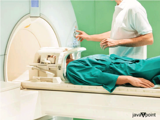
- Positioning: A mobile examination table that slides into the MRI scanner is used to situate the patient. To keep the patient still during the scan, the table could contain a headrest and straps. The technician will help the patient select a secure and comfortable position as proper placement is essential to getting reliable images.
- Coil Positioning: To enhance image quality, specialized coils are positioned all around the patient's head. These coils serve as receivers and send the MRI machine the signals the patient's brain emits so it can process them. To achieve the best signal detection, the technologist will carefully arrange the coils.
- Ear Protection: During the scanning procedure, MRI equipment makes loud banging noises. Earplugs or headphones are offered to the patient to shield their ears from the noise. Some facilities may provide headphones or music to aid in patient relaxation and anxiety reduction throughout the scan.
- Contrast Agent: To make particular brain disorders or structures more visible, a contrast agent may occasionally be given. Typically, an intravenous (IV) line is put into the patient's arm to accomplish this. In the generated images, the contrast agent's gadolinium-based component aids in highlighting particular brain regions.
- Communication: The technician will use an intercom system to speak with the patient after they are in a comfortable position. To address any worries or pain during the scan, it is crucial to establish clear communication. Additionally, the technologist will give directions on when it is okay to move and when to stay motionless.
- Scan Acquisition: The MRI machine comprises of a sizable cylindrical magnet with a patient-positioning bore in the middle. Protons in the patient's body are aligned by the machine's strong magnetic field. The protons then emit signals, which are picked up by the coils, as a result of the application of radio waves. A computer then creates cross-sectional scans of the brain using these inputs.
- Maintaining Stillness: To prevent image blurring during the scan, the patient must maintain complete stillness. Any movement could degrade the image's quality and require another scan. The technologist will keep an eye on the patient constantly from a nearby control room and offer direction or assurance when necessary.
- Multiple Sequences: To see distinct elements of the brain, MRI scans use several imaging sequences. Imaging techniques including T1-weighted, T2-weighted, and diffusion-weighted imaging are frequently used to assess various tissue properties like architecture, disease, and water diffusion. The relevant information may be captured by each sequence in a few minutes.
- Scan Completion: The technician will let the patient know the scan is finished once all necessary sequences have been acquired. The MRI table will slide out so the patient can adjust their posture and get hold of any personal items. The IV line will be cut if a contrast medication was given.
- Image Analysis: A radiologist, a specialist doctor trained in the interpretation of medical pictures, receives the obtained images. The radiologist will examine the pictures and produce a thorough report for the doctor. This report gives important details on the structure of the brain and any anomalies found during the scan.
How to Read Brain MRI?
The guidelines that help to understand the key components of a typical brain MRI report are:
- Technique: The report will start by outlining the precise MRI method utilized, including the magnetic field strength (such as 1.5 Tesla or 3 Tesla), the sequences used (such as T1-weighted, T2-weighted, and FLAIR), and any additional specialized sequences or contrast agents used (if relevant).
- General Findings: An overview of any notable discoveries made during the brain MRI is given in this section. It might contain details regarding the size and shape of the various brain organs, the existence of any masses or anomalies, and general observations regarding the cerebral cortex, ventricles, and supporting organs.
- Parenchymal Findings: The actual brain tissue is the main topic of this section. It could be used to indicate atrophy, abnormal signal intensities, or regions of anomalous enhancement (if contrast was utilized) in the grey matter (cortex) or white matter. Additionally, it might note whether the brain parenchyma has any infarcts (strokes), haemorrhages, or atypical lesions.
- Ventricular System: The ventricles, which are fluid-filled chambers in the brain, will be evaluated in the report. The ventricles' size and form will be discussed, and any enlargement (hydrocephalus) or obstruction to the passage of cerebrospinal fluid (CSF) will be searched for.
- Skull, Scalp, and Soft Tissues: The skull, scalp, and soft tissues that surround the brain are all examined in this area. Any anomalies or lesions in these regions, such as bone fractures, tumors, or infections, may be reported by it.
- Vascular Findings: The radiologist will assess the brain's blood vessels. This may entail checking the major veins and arteries for anomalies such as arteriovenous malformations (AVMs), aneurysms, or signs of ischemic alterations.
- Extra-Axial Structures: The structures that are present in the cranial vault but not in the brain tissue are examined in this section. It contains the meninges, which are the brain's protective coverings, as well as other parts including the pituitary and pineal glands. You may report any anomalies or masses in these locations.
- Impression/Conclusion: An overview and analysis of the results will be provided at the end of the report. The significance and potential clinical implications of the detected anomalies will be evaluated by the radiologist. If necessary, they might advise more diagnostic testing, follow-up imaging, or a specialist appointment.
Advantages and Disadvantages of Brain MRI
The advantages and disadvantages of brain MRI are:
Advantages:
- Non-invasive: The fact that brain MRI is a non-invasive method is one of its main benefits. MRI spares the patient from dangerous radiation exposure, unlike other imaging methods like CT scans that employ ionizing radiation. This makes it a safer choice, especially for people who may eventually need a lot of imaging tests.
- Detailed and Clear Images: MRI scans of the brain's architecture produce incredibly accurate and distinct images. Physicians can distinguish between distinct forms of brain tissue, such as grey matter, white matter, and cerebrospinal fluid, thanks to the superior soft tissue contrast it provides. This level of specificity is very helpful in the detection and follow-up of a variety of brain problems, including tumors, strokes, multiple sclerosis, and neurodegenerative diseases.
- Multiplanar Imaging: The brain can be imaged with MRI from many angles. It allows for the creation of pictures in the sagittal, coronal, and axial planes, which facilitates a thorough assessment of the various brain structures. This adaptability is especially useful for detecting lesions, and abnormalities, and assessing the connections between various brain regions.
- No Radiation Exposure: As was already explained, MRI doesn't use ionizing radiation. As a result, it is a good imaging technique for sensitive groups including kids and expectant mothers. There are no substantial health concerns associated with its repeated and cautious use.
Disadvantages:
- High Cost: Brain MRI scans are relatively pricey when compared to other imaging modalities since MRI equipment is expensive to buy and maintain. Particularly in nations with low healthcare resources or for people without proper insurance coverage, the high cost can be a substantial barrier.
- Long Scan Time: Brain scans with an MRI machine might take anywhere from 30 minutes to an hour or more to complete. Patients who have trouble staying motionless for long periods or who are claustrophobic may find the scan's duration to be unpleasant. But as technology has advanced, quicker imaging sequences have been created, sometimes cutting scan times.
- Contraindications and Limitations: Some people may not be able to undergo an MRI because they have specific metallic implants or gadgets inside of them. Metal objects may move or heat up due to the intense magnetic field, which could be harmful. The restricted environment within the MRI machine may also be difficult for patients with severe claustrophobia to bear.
- Noise and Discomfort: During the scanning procedure, MRI machines make loud pounding or thumping noises, which some patients may find unnerving. Although earplugs or headphones are frequently given to patients to lessen discomfort, some may still find the noise upsetting.
- Limited Availability: Compared to other imaging modalities like X-ray or CT scanners, MRI machines are not as commonly accessible. For patients who need urgent imaging, this limited availability may result in lengthier wait times, which could postpone diagnosis and treatment.
|

 For Videos Join Our Youtube Channel: Join Now


