| |

Brain ScanA brain scan is a term which is used to refer to a variety of imaging tests of our brain. Brain scans allow the doctor to "see inside" the skull without requiring surgery. Neurologists are very skilled in both performing and reading brain scans. 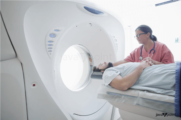
Why it's Necessary?A brain scan, as an imaging test, can assist the doctor in evaluating structures within the brain. A Positron Emission Tomography (PET) scan, for example, can tell the doctor that how well the human brain is performing its activity. A Neurologists will advise to perform brain scans to for diagnose or monitor neurological diseases or disorders in human brain. Brain scans are a great non-invasive way to determine neurological diagnoses. The data obtained from a brain scan will assist the doctor in treating your neurological issue. The doctor will propose a brain scan if there is reason to believe that someone has a neurological disorder or brain injury, or to rule out these conditions. Common Illnesses that are Treated by Brain ScansBrain scans can be used by doctors to diagnose and monitor a wide range of neurological diseases, including - Brain cysts or tumours, Anomalies of the blood vessels Multiple sclerosis (MS), Stroke Hydrocephalus or epilepsy Head trauma, including concussion. Consult with doctor if anyone have a neurological disorder that may necessitate monitoring with a brain scan. TypesDoctors are typically asked to do three types of brain scans: computed tomography (CT), magnetic resonance imaging (MRI), and positron emission tomography (PET). There are several subtypes of brain scans that can examine oxygen consumption in the brain, cerebral blood volume, and other aspects of brain structure or function. CT Scan of the BrainThis test produces images of organs, tissues, and bones by using ionising radiation (X-ray). CT scans are helpful because they can show the afflicted area of the brain and can be utilised immediately to detect bleeding or blockages. The operation can be performed with or without the addition of contrast dye, which highlights certain parts of the brain. In any event, you will be asked to change into a hospital gown and lie down on a special table that fits through the tight access to the imaging equipment. You will be given an intravenous (IV) line if iodine-containing contrast dye is utilised. The entire procedure takes roughly 20 minutes. CT scans are used to diagnose the following conditions: brain injury, bone or vascular abnormalities, and hydrocephalus (a buildup of brain fluid). MRI Brain ScanMRI, unlike CT scans, which use ionising radiation to produce images, employs a magnetic field and radio waves. You will be instructed to strip naked and lie on a table that will be pushed through the magnetic resonance imaging machine. During an MRI, you cannot have any magnetic metal on or within your body. Gadolinium is the most often used contrast dye for brain MRI; if it is utilised, you will be given an IV line. A detector is also put over the head for brain MRIs. Tissue connection, Blood flow measurements, Mineral deposit information, Diagnosis of stroke, multiple sclerosis, traumatic brain injury, tumours, infection, inflammation, and other issues with are diagnosed by MRI. MRIs are painless and risk-free, though claustrophobic or obese patients may suffer discomfort. MRIs usually take one hour to complete. Functional MRIs (fMRI) produce real-time pictures of blood flow in the brain. This is very useful for identifying active locations. They can also be utilised to identify brain regions involved in motor function, language, or emotion. They are also useful in the research of degenerative illnesses. PET Scan of the BrainWhile CT and MRI scans examine brain structure, PET scans examine brain function. This test involves the infusion of a radioactive tracer material into your hand or arm via an IV line. It takes roughly an hour for this stuff to circulate through your system. Following that, you will be placed on a table and scanned by a machine. PET scans employ gamma radiation. PET scans detect tumours and damaged tissue, as well as blood flow and tissue metabolism. They are particularly useful in assessing patients with epilepsy or memory issues. PET scans can detect cancer and assess how well you are responding to treatment. PET scans might last anywhere between 30 minutes and two hours. Some patients have difficulty lying motionless for long periods of time, while others are apprehensive and uneasy in the enclosed atmosphere of the scanning equipment. If you are claustrophobic, ask to your doctor or technician about how you may make the scan more comfortable for you. RisksBrain scans have few dangers in general. The radiation used in CT and PET scans is extremely low and does not stay in your body for long. Pregnant women are at a higher risk because foetuses are more sensitive to the effects of radiation. For pregnant women, MRIs are a superior alternative. Before having a diagnostic scan or other procedure, please see your doctor if you are pregnant or suspect you are pregnant. An allergic reaction to the radioactive dye is also possible. This is extremely unusual, but it might cause swelling, pain, or redness at the injection site.
Next TopicCerebrovascular Accident
|
 For Videos Join Our Youtube Channel: Join Now
Feedback
- Send your Feedback to [email protected]
Help Others, Please Share


