| |

Human Brain Weight in KgAnatomy, biological anthropology, animal science, and evolution often discuss the size of the brain as a research issue. MRI scans, or the skull's volume, are two methods for determining brain size. Analyzing the volumetric measures of the brain may be done using neuroimaging intelligence tests. The relationship between brain size and IQ has received much attention in the field of "intelligence testing." It will be discussed more in the section on intelligence since this issue is debatable. For all animals, including humans, the measurement of cranial capacity and brain size is crucial. Human Brain SizeWhile the cerebellar hemispheres are usually closer in size, the right cerebral hemisphere is usually bigger than the left in humans. About 1.5 kg (3.3 lb) is the typical weight of an adult human brain. About 1370 g on average weighs males, and 1200 g on average weighs women. Despite significant individual variation, the volume is around 1260 cm3 for males and 1130 cm3 for women. Another research said that a newborn human's brain weighs between 350 and 400g while that of an adult is between 1,300 and 1,400g. 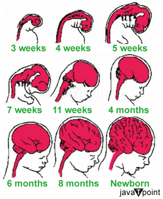
In contrast to body mass, where one can depend on a single value as being unambiguously accurate, there is a range of volume and weights. Furthermore, as individual variation is often far less significant than species-level variation, it is crucial to remember that it is more relevant. Intraspecific and interspecific variations have different processes. Variation and Evolution of the Human BrainExcept for the extinct Neanderthals, whose brain size was greater than that of contemporary Homo sapiens, brain size increased from early primates through hominids and eventually to Homo sapiens. The human brain has grown in size during human evolution (see Homininae), from around 600 cm3 in Homo habilis to 1680 cm3 in Homo neanderthalensis, the hominid with the largest brain. Research that concluded that the drop "was surprisingly recent, occurring in the last 3,000 years" is one example of evidence suggesting that the average brain size has shrunk since that time. The finding was reached using datasets that are too different to allow for a quantitative comparison, according to a reanalysis of the same data, which indicates that brain size has not decreased. The gene mutation that results in microcephaly, a neural developmental condition that affects cerebral cortical volume, is highlighted by proponents of recent increases in brain size. Similar to this, sociocultural arguments highlight the externalization of knowledge and collective decision-making as potential reasons, pointing to the emergence of social systems of dispersed cognition, social organization, labor division, and information exchange. Homo floresiensis is a hominid that has remains from the Indonesian island of Flores that date from 60,000 to 100,000 years ago. While Homo habilis is thought to have gone extinct much earlier (about 1.65 million years ago), despite its relatively derived position in the human lineage, CT imaging of its skull revealed that its brain capacity was only 417 cm3, less than even that of Homo habilis. It is thought that island syndrome, in which insulating species' brains shrink due to decreased predation danger, is to blame for this decline in brain size. This lowers the baseline metabolic rate without significantly raising the danger of predation, which is advantageous. It has been suggested that even with extremely tiny brains, very high levels of intellect and reasonably normal functioning are conceivable by looking at exceptional examples of hydrocephalus, such as those described by John Lorber in 1980 and by rat research. It is unknown what inferences concerning, for example, brain capacity, redundancy, mechanics, and size needs may be made from such accounts. Biogeographic VariationThe search for racial or ethnic differences in brain size is often seen as a pseudoscientific endeavor historically connected to scientific racism and efforts to establish a race-intellectual hierarchy. Instead of using direct brain observations, most attempts to prove this have relied on indirect data that evaluated measures of the skull. These are regarded as being disproven scientifically. We find little evidence to support the use of brain size in taxonomic evaluation (other than with paleontological extremes over time), according to a large-scale 1984 study of global variation in skull sizes. This study found that variation in skull and head sizes is unrelated to race but to climatic heat preservation. Human Brain Variation According to the SexThe typical human baby's brain measures 369 cm3 at birth and grows to around 961 cm3 in the first year of life before beginning to shrink. Teenage years are when brain capacity peaks, and after age 40, it starts to decline at a rate of 5% each decade, picking up pace around age 70. An adult male's brain weighs an average of 1,345 grams (47.4 oz), while an adult woman's brain weighs an average of 1,222 grams (43.1 oz). Men typically have bigger bodies than women. However, this does not account for neuron density or the ratio of the mass of the brain to that of the body. It has been shown that males often have larger cerebral, cerebellar, and cerebral cortical lobar volumes, except for the left parietal. More specialized brain areas vary by gender variations in size. According to studies, males tend to have a bigger amygdala and hypothalamus than women, and vice versa for the caudate and hippocampi. Kelly (2007) found that compared to intracranial volume, height, and weight, males had a larger proportion of white matter and cerebrospinal fluid, while women had a higher percentage of grey matter. These studies do show substantial inter-individual variation. 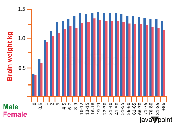
However, Yaki (2011) observed no statistically significant gender differences in the grey matter ratio in the sample of 758 women and 702 men aged 20-69, except in the third and sixth decades of life. The grey matter ratio of the typical man in their third decade (ages 20-29) was substantially greater than that of the typical female in the same age group. In contrast, the typical woman had a considerably higher grey matter ratio among participants in their sixth decade of life. At the same time, there was no appreciable difference among those in their seventh decade of life. While white matter and ventricular sizes rise, the total cerebral and grey matter volumes peak between 10 and 20 years (earlier in females than in boys). The brain growth process often follows a pattern of childhood peaks and teenage falls (e.g., synaptic pruning). Boys' average brain volume is almost 10% greater than girls', which aligns with adult results. The high variability of brain size, even in tightly defined groups, for example, children at the same age may have as much as a 50% difference in total brain volume, is noteworthy. However, such differences should not be interpreted as imparting any functional advantage or disadvantage. Gross structural measures may not reflect functionally relevant factors such as neuronal connectivity and receptor density. The hippocampus volume of young females is typically greater than that of boys, although the amygdalae are bigger in boys. However, several studies have shown that males had larger synaptic densities. For example, a 2008 research indicated that men had an average synaptic density of 12.9 × 10⁸ per cubic millimeter, compared to 8.6 × 10⁸ per cubic millimeter in women, a 33% difference. Other studies have confirmed this discrepancy, finding an average of 4 billion more neurons in the male brain, with an average of 7,000 synaptic connections between each neuron. The brain structure undergoes significant dynamic changes through maturity and aging, with significant individual diversity. Men have larger volume loss in the frontal, temporal, and entire brain volumes as they age, while women experience greater volume loss in the hippocampi and parietal lobes. The grey matter volume reduction is more pronounced in men, although it varies by location in both sexes, with certain regions showing little to no age impact. Although there is heterogeneity within brain areas, the overall white matter volume does not seem to decrease with age. Genetic ContributionAccording to adult twin studies, the adult brain's total size is highly heritable (between 66% and 97%). Regionally, the impact varies throughout the brain, with strong frontal lobe heritabilities (90-95%), intermediate hippocampi estimates (40-69%), and environmental influences on various medial brain regions. Furthermore, environmental influences are the primary explanation for the lateral ventricle volume, indicating that these factors also affect the brain tissue around the lateral ventricle. Brain anatomy and cognitive abilities may be linked through genes, or the latter may have a lifelong impact on the former. Numerous candidate genes have been found or proposed. However, these need to be replicated.
Next TopicLargest Part of Brain
|
 For Videos Join Our Youtube Channel: Join Now
Feedback
- Send your Feedback to [email protected]
Help Others, Please Share


