| |

Brain FeverThe medical condition known as "Brain Fever" causes one region of the brain to swell up and develop symptoms like a fever. The term is archaic and most frequently used in Victorian literature, where it usually refers to a potentially fatal sickness brought on by a great emotional trauma. 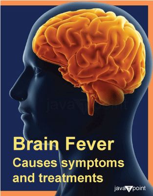
Different conditions of Brain FeverThe following conditions could be characterised as brain fever:
1. EncephalitisEncephalitis, or inflammation of the brain, can be brought on by a virus, bacteria, medication, or immune system issue. Encephalitis is a rare but frequently serious illness that requires immediate medical care. In what ways can Encephalitis influence the body?Fever, headaches, and neck pain are physical indicators of encephalitis. Additionally, it can influence how the brain (cognitive) functions, which can cause confusion and alter behaviour. Sometimes, cognitive encephalitis symptoms continue long after the physical ones have subsided. CausesDepending on the reason, you may develop a certain type of encephalitis. There are also several causes, such as:
In rare cases, parasites and bacteria can both cause bacterial encephalitis. What signs of Encephalitis?You'll probably have neurological and bodily problems. Some physical signs include:
Symptoms of neurologic encephalitis include:
Tests and DiagnosisYour chances of recovering are better the earlier you obtain care. Your first course of action for mild encephalitis symptoms may be to visit an urgent care facility. The best place to receive care for serious conditions, such as seizures and loss of consciousness, is an emergency department. Which tests would require?
You may need the following tests:
Control and TreatmentDepending on the kind of encephalitis you can get to know how serious it is, and what type of treatment you may require for a it. You may require:
How quickly will it recover?Treatments for viral and autoimmune encephalitis may start to act as soon as a few days later. However, the brain can take some time to heal. Some people experience chronic cognitive deficits that necessitate rehabilitative treatments and dietary changes. 2. MeningitisMeningitis is an infection of the tissues lining the brain and spinal cord. Infection is typically to blame for it. It may be lethal, thus quick medical attention is necessary. Numerous types of bacteria, viruses, fungi, and parasites can cause meningitis. Many illnesses are communicable from person to person. A tiny percentage of cases are brought on by accidents, cancer, and medicines. The deadliest variety of meningitis, caused by bacteria, has a 24-hour mortality rate. Any age group might be affected by meningitis. To prevent some of the major bacterial causes of meningitis, there are both effective therapies and vaccines. Meningitis, however, continues to pose a serious hazard on a global scale. Acute bacterial meningitis can be brought on by four primary factors:
These bacteria also cause other serious illnesses like sepsis and pneumonia in addition to being the cause of more than half of meningitis deaths worldwide. Meningitis can also be brought on by other bacteria like Mycobacterium tuberculosis, Salmonella, Listeria, Streptococcus, and Staphylococcus, viruses like enteroviruses and the mumps, fungi, like Cryptococcus, and parasites like Amoeba. Who is in danger?Meningitis can affect people of any age, but small children are the most vulnerable. Newborns are most at risk from Group B streptococcus, while young children are most at risk from meningococcus, pneumococcus, and Haemophilus influenzae. Meningococcal disease has a specific risk to adolescents and young adults, whereas pneumococcal disease poses a special risk to the elderly. Meningitis is a threat to people everywhere. The sub-Saharan African region known as the African Meningitis Belt, which is particularly identified to be at high risk of outbreaks of meningococcal but also pneumococcal meningitis, has the largest disease burden. The danger is higher when people are living near to one another, such as during big gatherings, in refugee camps, in crowded dwellings, or in school, military, and other workplace environments. A variety of meningitis forms can also be increased by immune weaknesses including HIV infection or complement insufficiency, immunosuppression, and active or passive smoking. TransmissionThe mode of transmission differs by organism. Most meningitis-causing bacteria, including meningococcus, pneumococcus, and Haemophilus influenzae, reside in the human nose and throat. By way of throat secretions or respiratory droplets, they pass from person to person. Humans frequently have Group B streptococcus in their vagina or stomach, and it can pass from mother to kid around the time of birth. Although carrying these organisms is mostly safe and aids in the development of antibody to infection, the bacteria can occasionally enter the body and cause meningitis and sepsis. Symptoms and SignsA patient's clinical characteristics may vary depending on the cause, disease course (acute, subacute, or chronic), brain involvement (meningo-encephalitis), and systemic effects (such as sepsis).
Similar signs and symptoms can be found in meningitis caused by bacteria or viruses. Some forms of meningitis may have more severe symptoms than others, necessitating a particular course of therapy. Other signs of bloodstream infections (septicaemia), which can swiftly progress to sepsis due to bacterial meningitis pathogens, include:
One in five individuals who survive a case of bacterial meningitis may experience long-term complications. These side effects include scarring and limb amputations following infection, as well as hearing loss, seizures, limb weakness, problems with vision, speech, language, memory, and communication. PreventionThe best defence against prevalent forms of bacterial meningitis is vaccination. The following can induce meningitis:
Meningitis caused by viruses and bacteria can transmit from person to person. In the case of bacterial meningitis, discuss taking antibiotics with your doctor or nurse.
1. Vaccination Meningococcal, pneumococcal, and Haemophilus influenzae illness vaccines have been approved for use for many years. There are numerous strains of these germs (referred to as serotypes or serogroups), and vaccines are made to guard against the most dangerous ones. There is no all-purpose vaccination. Meningococcus serogroup A was responsible for 80-85% of meningitis epidemics in the African meningitis belt before the meningococcal A conjugate vaccine was introduced through widespread preventive campaigns (from 2010) and routine immunisation programmes (since 2016). To prevent the return of epidemics, introduction into routine immunisation campaigns must continue, and high coverage must be maintained. 2. Preventative Antibiotics (Chemoprophylaxis) When promptly administered, antibiotics for those near those who have meningococcal disease reduce the chance of transmission. Chemoprophylaxis is advised for close family contacts outside the African meningitis area. Chemoprophylaxis for close contacts is advised in non-epidemic circumstances within the meningitis belt. The recommended antibiotic is ciprofloxacin, though ceftriaxone is an option. In many nations, it is advised to identify women whose offspring are at risk of contracting group B streptococcal illness. In order to keep babies from contracting Group B streptococcal infection during delivery, at-risk mothers are given intravenous penicillin. DiagnosisA clinical examination is the initial stage in the diagnosis of meningitis, which is then followed by a lumbar puncture. When the spinal fluid is examined under a microscope, the bacteria are occasionally visible. To support or confirm the diagnosis, the bacteria are grown from blood or cerebrospinal fluid samples, quick diagnostic tests are run, or Polymerase Chain Reaction (PCR) is employed. To determine control methods, it is critical to identify serogroups and antibiotic susceptibility. Whole genome sequencing and molecular typing reveal additional strain distinctions and guide public health interventions. 2. CerebritisThe inflammation of the cerebrum, which is responsible for several crucial processes like speech and memory, is known as cerebritis. On CT scans, it is additionally described as a purulent nonencapsulated parenchymal infection of the brain (ill-defined low-density area with peripheral enhancement) that is consistently difficult to distinguish from neoplasms. In most cases, cerebritis results from an underlying disease that inflames the brain tissue. It is frequently discovered in lupus patients. Both adults and children are susceptible to lupus cerebri. The time that the central nervous system is affected might range from a few minutes, as in a transient ischemia attack or a classic migraine, to years, as in dementia. Neurological impairments that ensue may occasionally be fatal and be either temporary or permanent. SymptomsCerebritis symptoms can range from minor to severe. The degree of oedema and the level of raised intracranial pressure both influences how severe the symptoms are. Headaches, sadness, worry, and memory loss are examples of mild symptoms. When the brain or nervous system is attacked due to immune system issues, inflammation of the brain may occasionally be present. Inflammation can lead to significant issues such headaches, seizures, vision issues, vertigo, behavioural abnormalities, and even stroke. Severe lupus cerebritis symptoms include psychosis, dementia, peripheral neuropathy, cerebellar ataxia (loss of muscular coordination, usually on one side of the body), and chorea (jerky, uncontrollable movements). Patients with systemic lupus have a 3-20% stroke incidence, with the risk being highest in the first five years of the illness. More than 20% of people with systemic lupus experience peripheral neuropathy, such as carpal tunnel syndrome, and 10-15% experience cranial nerve palsies. CausesSystemic Lupus Erythematosus (SLE) is one of the most frequent causes of cerebritis, with more than half of American lupus patients believed to have some kind of lupus cerebritis. It is uncertain what pathophysiological mechanism causes lupus cerebritis. The onslaught of numerous autoimmune system alterations, including the following, is likely what causes the suggested mechanisms: immunological circulating complexes. The blood-brain barrier is broken down because of the immune complexes, which are made up of DNA and anti-DNA. Upon autopsy, these circulating complexes were discovered entrapped in the very vascular choroid plexus of SLE patients. However, only 10% of patients with cerebral lupus have true vasculitis.
But it's unclear which pathway in lupus patients causes cerebritis. It's possible for all mechanisms to operate simultaneously or separately, according to experts. Cerebritis may, in extremely rare circumstances, arise from a Klebsiella pneumoniae infection. An infection brought on by bacteria, viruses, or other living things is another factor that might lead to cerebritis. Infections can develop as a result of trauma or when infectious substances reach the brain through the sinuses. Even though the body has developed defences that are especially intended to stop this, some infections are still capable of breaching the blood-brain barrier and accessing the brain through the bloodstream. TreatmentThere is currently no treatment for lupus. However, autoimmune activity is suppressed to cure lupus cerebritis. When infections are the underlying cause, medication is utilised as treatment, with the goal of primarily curing the illness. Steroids can be used to reduce oedema caused by inflammation. Surgery can be required to release the pressure on the brain if the swelling seems to have reached a worrisome degree. The development of an abscess necessitates surgery as well since the abscess must be drained. 4. Scarlet FeverScarlatina, also referred to as scarlet fever, is a type of Group A streptococcal infection (Group A strep) brought on by the GAS Streptococcus pyogenes. Children between the ages of five and fifteen are typically affected. Some of the warning signs and symptoms include a distinctive rash, swollen lymph nodes, fever, headache, and sore throat. The rash is often itchy and feels like sandpaper, and the face is flushed. A rough, crimson tongue is another possibility. Exotoxins cause capillary injury that results in the rash. Sneezing or coughing is usually how the bacterium is spread by people. Additionally, touching your lips or nose after touching something with the bacteria on it can spread it. Swabs of the throat are often cultured to confirm the diagnosis. Few persons with strep throat or streptococcal skin infections go on to develop scarlet fever. Antibiotics can be used to treat the condition since they stop the spread of the illness' symptoms and most consequences. If treated, scarlet fever usually has positive outcomes. Ridiculous rheumatic fever, arthritis, and renal damage are long-term effects of scarlet fever. Scarlet fever is not preventable by a vaccination. The severity of the illness was already decreasing even before the Second World War and the discovery of antibiotics, even though it was a leading cause of death for children in the early 20th century. This may be due to better living conditions, the introduction of better control measures, or a decrease in the virulence of the bacteria. In recent years, there have been indications of antibiotic resistance; there were outbreaks in Hong Kong in 2011 and the UK in 2014, as well as the occurrence of the diarrhoea. According to research presented in October 2020, the bacterium has developed more deadly variants because of three viruses infecting it. SymptomsScarlet fever often appears after a group of a streptococcal infection, such as streptococcal tonsillitis or more frequently streptococcal pharyngitis, that results in strep throat and typically starts with a sore throat, fever, and malaise. The presence of a headache, nausea, vomiting, and stomach pain is also possible. Pharyngotonsillitis is a common symptom of these illnesses. The characteristic widespread rash then follows the strep throat signs and symptoms, which are those of a strep throat. The rash often occurs one to two days after feeling sick, but it can also show up earlier or up to seven days later. It typically hurts to swallow. Nevertheless, not all instances start with a fever, the degree of exhaustion can differ, sore throats and tongue alterations can be minor or non-existent, and some cases may start with a patchy rash rather than a widespread one. The Mouth and ThroatStreptococcal pharyngitis in a child's throat with patches on the roof of the mouth and a red throat after a positive throat culture. Exudate frequently covers the tonsils, which can sometimes seem red and bloated. The roof of the mouth may have a few little red patches and the throat may be inflamed. The uvula may appear enlarged and inflamed. Neck lymph nodes that are swollen and painful are linked to between 30% and 60% of cases. The tongue may have a creamy coating during the first two days of illness, from which red, swollen papillae protrude. RashScarlet fever rash on dark skin scarlet fever rash on light skin red cheeks and a pale mouth area in scarlet fever Typical scarlet fever red cheeks and rash. The distinctive rash is known as "scarlatiniform" and manifests as a generalised skin redness with little goosebump-like lumps. Before expanding into tiny bumps that extend to the arms and legs, it often starts off as small flat areas on the neck or torso. It often has a sandpaper-like roughness to it. There may be a pale region around the mouth and flushed cheeks. On white and pale skin, the scarlet fever rash normally appears red. On brown or black skin, however, the bumps are typically larger, the skin feels less like sandpaper, and the perioral pallor is less noticeable. The soles and palms are unaffected. When pressure is applied, the reddish skin blanches. Although the skin may itch, it is not unpleasant. The inside of skin folds and crevices may have a more pronounced redness. The armpits and elbow pits include these lines of petechiae, which appear as pink or red regions. The primary rash takes about a week to go away. The skin of the usual fingers and toes may peel for several weeks after this. Desquamation typically starts on the face and works its way down the body. This peeling may occasionally be the only indication that someone has scarlet fever. If the scarlet fever case is straightforward, recovery from the fever and clinical symptoms-aside from the desquamation process-occurs in 5-10 days. Desquamation will leave the skin with a burnt appearance. ComplicationsScarlet fever complications can be categorised as either suppurative or nonsuppurative-
TreatmentThe cornerstone of scarlet fever treatment is the use of antibiotics to fight the streptococcal infection. To reduce the length of an infection, antibiotics should be given as soon as feasible. Preventing the kid from developing a suppurative or nonsuppurative consequence, particularly acute rheumatic fever, is one of the key goals of treatment. The development of acute rheumatic fever in the youngster is quite rare if antibiotics are started within nine days. Even with treatment, the skin's outer layer will eventually peel off. After taking antibiotics for 24 hours, a child is no longer contagious (unable to spread the virus to another child). During the first two weeks, an infected person is most likely to transmit the infection to another person. Penicillin V, which is used orally, is the preferred antibiotic. Children who cannot swallow tablets can be given amoxicillin, which is available in a liquid version and is similarly effective, in nations lacking a liquid Penicillin V product. A 10-day therapy regimen is advised. If taking pills is not an option, benzathine penicillin G can be injected intramuscularly once. A first-generation cephalosporin is used if the patient is allergic to the class of antibiotics (beta-lactam antibiotics) that both penicillin and amoxicillin belong to. Cephalosporin antibiotics, however, can still have negative effects in patients who have had Clindamycin or erythromycin should be used in certain situations instead of tonsillectomy, which was once a viable option for treating recurrent streptococcal pharyngitis because a person can still contract group.
Next TopicBrain Fog Meaning
|
 For Videos Join Our Youtube Channel: Join Now
Feedback
- Send your Feedback to [email protected]
Help Others, Please Share


