| |

Brain StructureThe brain has a complex and mind-boggling structure. It is a magnificent organ that controls our ideas, emotions, and behaviour. The brain is a complicated network made up of billions of linked neurons that controls how information is processed and how our whole life is orchestrated. Investigating the amazing structure of the brain gives a fascinating look at how our cognitive abilities function inside our brains and offers important insights into various neurological illnesses. We explore the main components of brain structure in this article, revealing how complexly they are organized and emphasizing the importance of comprehending this miracle of nature. 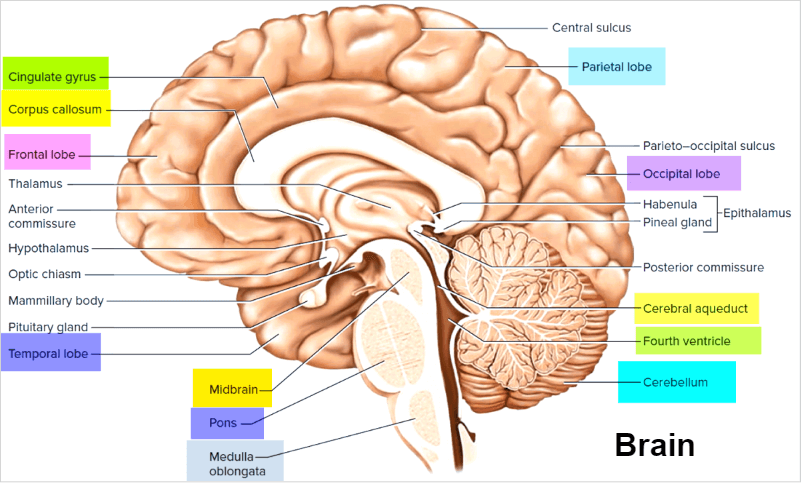
Anatomy of the BrainA miraculous three-pound organ, the brain directs all bodily processes, deciphers data from the outside world, and embodies the spirit and mind. Intelligence, creativity, emotion, and memory are just a few functions the brain regulates. The brain comprises the cerebellum, brainstem, and cerebrum and is protected by the skull. The five senses that we have-sight, smell, touch, taste, and hearing-all send information to the brain, sometimes in large quantities. It organizes the signals into a coherent whole that makes sense to us and is easy to remember. Thoughts, speech, memory, arm and leg movement, as well as the operation of several bodily organs, are all controlled by the brain. The central nervous system (CNS) is made up of the spinal cord and brain. The peripheral nervous system (PNS) is made up of cranial nerves that come from the brain and spinal nerves that come from the spinal cord. From infancy through maturity, the brain's weight fluctuates. The typical brain weighs around one pound at birth, and it increases to roughly two pounds during youth. The typical adult female brain weighs around 2.7 pounds, and the average adult male brain weighs about 3 pounds. Main Components of BrainThe cerebrum, cerebellum, and brainstem make up the brain. 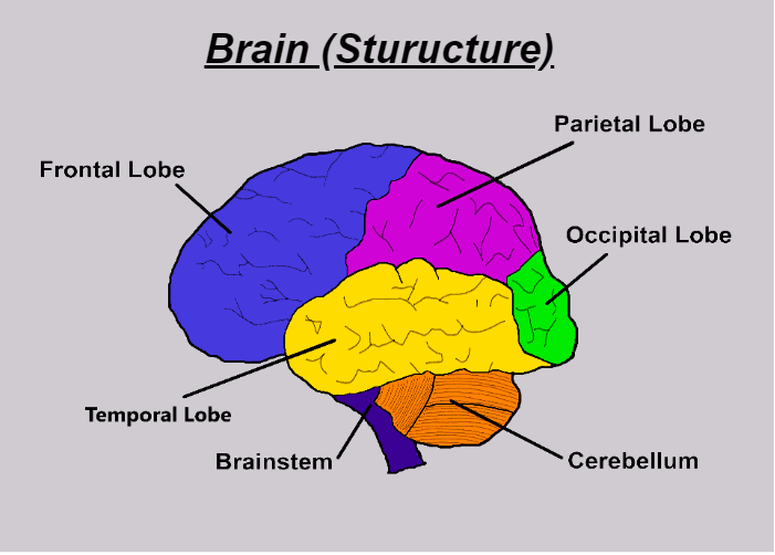
Right and left hemispheres make up the cerebrum, the brain's principal structural component. It carries out higher cognitive tasks, including speaking, thinking, emotions, learning, fine motor control, and processing touch, vision, and hearing. The cerebellum is located just below the skull. Its major goal is to keep your muscles coordinated, balanced, and seated. The spinal cord, brain, and cerebellum communicate with one another through the brainstem. Aside from breathing, it also automatically regulates other bodily functions like digestion, heart rate, body temperature, waking and sleeping patterns, and sneezing, coughing, vomiting, and swallowing. The two halves of the Cerebrum - Right Brain vs Left BrainThe right and left hemispheres make up the cerebrum's two halves. The corpus callosum, which connects them, is a collection of fibres that carries signals from one side to the other. Each hemisphere controls the body's opposing side. Your left arm or leg might be weak or paralyzed due to a stroke affecting your brain's right side. Each hemisphere performs different tasks. Speech, understanding, mathematics, and writing are generally under the authority of the left hemisphere. The right hemisphere governs creative, spatial, artistic, and musical abilities. About 92% of people use their left hemisphere more often than their right while speaking and writing. Brain LobesThe different fissures that separate the cerebral hemispheres create lobe-like divisions in the brain. Each of the four hemispheres comprises the frontal, temporal, parietal, and occipital lobes. Again, distinct regions inside each lobe may be identified that carry out very particular tasks. Knowing that the different lobes of the brain work in conjunction is important. The relationships between the brain's lobes and between its right and left hemispheres are exceedingly complex. Frontal LobesThe frontal lobes are the largest and carry out the most functions among the four lobes. These include cognitive, behavioural, and intellectual processes and speech and voluntary motor abilities. The primary motor cortex, also known as the precentral gyrus, contains the regions responsible for producing movement in different body portions. The prefrontal cortex greatly impacts how we remember things, think, focus, behave, and feel. The premotor cortex refers to the region adjacent to the primary motor cortex. It guides eye and head movements as well as one's sense of orientation. The Broca region of the frontal lobe is frequently on the left side and is essential for language development. Occipital LobesThe lobes at the back of the brain allow humans to receive and process visual information. They alter how humans view structure and colour. The right occipital lobe processes visual information from the left visual area, whereas the left occipital lobe does the same for the suitable visual space. Parietal LobesThese lobes interpret signals from the motor, sensory, visual, audible, and memory regions of the brain. Objects get significance based on a person's recollection, memory and newly acquired sensory information. Temporal LobesThese lobes may be separated into two halves and found on either side of the brain around the ear level. The ventral (base) and lateral (side) surfaces comprise each hemisphere, which contains two portions altogether. Visual memory is aided by a region on the right side of the brain, which also aids with facial and object recognition. A region on the left side of the brain aids people's ability to retain and comprehend language. LanguageThe left hemisphere of the brain, commonly known as the "dominant" hemisphere, is usually responsible for controlling language and speech. The right hemisphere's primary functions include interpreting visual information and spatial processing. Roughly one-third of left-handed people's speech functions may be on the right side of the brain. Left-handed people may need additional testing before any procedure in that area to determine whether their speech centre is on the left or right side. A brain injury, most usually a stroke or other traumatic event, can produce aphasia, a linguistic problem. It has an impact on writing, reading, speaking, and comprehension. The injured brain region determines the form of aphasia. The left frontal lobe is where Broca's area is located. If this region is affected, it could be difficult to move the tongue or the muscles in the face to make speaking sounds. Broca's aphasia refers to a condition in which a person may still read and understand the spoken language but finds it difficult to speak and write (i.e., struggles to form letters and words and scribbles outside of lines). In the left temporal lobe is a region known as Wernicke's area. Wernicke's aphasia results from damage to this region. The speaker could use lengthy, meaningless phrases, ad hoc word combinations, and other verbal tics. They may have the ability to speak, but since they have trouble interpreting speech, they are unable to correct their errors. The Surface of the Cerebrum: CortexThe cortex is the term for the cerebrum's exterior portion. It has a folded look with hills and valleys. The cortex typically contains about 16 billion neurons with the cerebellum containing an additional 70 billion, for a total of 86 billion; all of them are grouped into several layers. The grey-brown colour of the cortex, caused by the nerve cell bodies, gives it its name, i.e., gray matter. Long nerve fibres (axons), also known as white matter, go underneath the cortex and link various brain parts together. The cortex folds, increasing the brain's surface area and allowing more neurons to fit inside the skull, providing larger functions. Each fold is referred to as a gyrus, and each groove between folds is a sulcus. The folds and grooves that help identify particular parts of the brain have specific names as well.
Protection Layer of Brain - MeningesThree layers of tissue known as meninges surround and safeguard the brain and spinal cord. The dura mater, arachnoid mater, and pia mater are the three layers going inward from the outermost. The meningeal and periosteal layers of the dura mater, which tightly borders the inside of the skull, are joined and only physically separate to produce the venous sinuses. The dura makes little folds or chambers. The falx and the tentorium are two distinctive dural folds. The tentorium and falx divide the cerebrum from the cerebellum and the right and left hemispheres of the brain, respectively. A thin, web-like membrane called the arachnoid mater surrounds the whole brain. Elastic tissue makes up the arachnoid. The Pia mater follows the folds and grooves of the brain's surface. Numerous blood arteries in the pia mater extend far into the brain. The subarachnoid space is the region between the arachnoid and the pia. The cerebrospinal fluid flows and supports the brain in this region. Cells of the BrainNeurons (nerve cells) and glia cells are the two different types of cells that make up the brain, such as: Nerve CellsNeurons come in a variety of sizes and forms, but they all have a cell body, dendrites, and an axon. The neuron sends information through chemical and electrical impulses. Think about the wiring in your house. A light bulb will shine when a light switch is turned on thanks to the multiple wires that make up an electrical circuit. Likewise, when stimulated, a neuron will transfer its energy to nearby neurons. A synapse is a very small opening or gap via which neurons "talk" to one another. The many dendrites that each individual neuron possesses allow it to take up signals from other nerve cells like an antenna. The cell body receives these messages and decides whether to send them on or not. Significant messages are sent at the axon's tip, where neurotransmitter-containing sacs enter into the synapse. In order to trigger the receiving nerve cell to deliver the message, the neurotransmitter molecules must cross the synapse and fit into certain receptors. Glia CellsThe brain's glia cells, named after the Greek word for glue, typically help feed, shield, and support neurons structurally. Glia is the most prevalent type of cell in brain tumours and has between 10 and 50 times more of them than nerve cells. The guardians, called astrocytes, control the blood-brain barrier, letting chemicals and nutrients to communicate with neurons. They regulate electrical impulses, homeostasis, neural defence, and repair. Myelin, a fatty material made by oligodendroglia cells, insulates axons and speeds up the transmission of electrical signals. Ependymal cells are responsible for lining the ventricles and secreting cerebrospinal fluid (CSF). Immune cells called microglia defend the brain from pathogens and remove waste. Additionally, these cells also prune synapses. Cerebrospinal Fluid and VentriclesThe ventricles are hollow spaces in the brain that are filled with fluid. The choroid plexus, which resembles ribbon-like structures and is located inside the ventricles, is responsible for producing clear, colourless cerebrospinal fluid (CSF). CSF circulates inside and around them to protect the brain and spinal cord from damage. This fluid that circulates is continuously absorbed and refilled. The lateral ventricles are two large ventricles located deep inside the cerebral hemispheres. A different hole known as the foramen of Monro connects each of them to the third ventricle. A long, thin tube known as the aqueduct of Sylvius joins the third ventricle with the fourth ventricle. The CSF enters the subarachnoid area through the fourth ventricle, bathes, and supports the brain there. Arachnoid villi, specialized cells located in the superior sagittal sinus, recycle (or absorb) CSF. The amount of CSF generated and absorbed is kept in balance with one another. A breakdown or obstruction in the system may result in a buildup of CSF, which may enlarge the ventricles (hydrocephalus) or result in a buildup of fluid in the spinal column (syringomyelia). Blood FlowThe internal carotid arteries and the vertebral arteries, which are two paired arteries, provide blood to the brain. The internal carotid arteries fuel the majority of the cerebrum. The cerebellum, brainstem, and base of the cerebrum are all supplied by the vertebral arteries. The basilar artery is created when the right and left vertebral arteries combine after passing through the skull. At the Circle of Willis, located at the brain's base, the basilar artery and the internal carotid arteries "communicate" with one another. A key safeguard of the brain is the link between the vertebral-basilar and internal carotid systems. Collateral blood flow may reach the Circle of Willis and stop brain injury if one of the primary arteries becomes blocked. The brain has a vastly distinct venous circulation from the rest of the body. Typically, arteries and veins flow side by side as they nourish and drain particular bodily parts. So, it stands to reason that there would be internal carotid veins in addition to a pair of vertebral veins. The brain, however, operates differently. Venous sinuses, which should not be confused with the air sinuses in the face and nasal area, are formed by the integration of the main vein collectors into the dura. The internal jugular veins carry brain blood to the venous sinuses from where it is taken. The superior and inferior sagittal sinuses drain the cerebrum, while the cavernous sinuses drain the anterior skull base. The MemoryEncoding (choosing which information is relevant), storing, and remembering are the three stages of the complicated process of memory. Various brain regions process different forms of memory. Encoding is the process through which an experience is transferred from short-term to long-term memory in your brain. The prefrontal cortex is where short-term memory, often working memory, takes place. It can only hold roughly seven items at a time and only keeps data for about a minute. You may use it, for instance, to call a phone number that someone just gave you. Additionally, it interjects throughout the reading to help you remember the previous line so that the subsequent one makes sense. When you wish to remember anything for a longer period of time, your long-term memory-which is processed in the hippocampus of the temporal lobe-is triggered. The capacity of this memory is infinite in both content and duration. Along with facts and data, it usually includes personal memories. The cerebellum processes memory for skills before sending information to the basal ganglia. It keeps track of learned automatic skills like tying shoes, playing an instrument, and riding a bike. SkullThe skull is made of bone tissue to safeguard the brain from harm. About 8 bones join together along suture lines to create the skull. The frontal, parietal (2), temporal (2), sphenoid, occipital, and ethmoid bones are a part of this group. The 14 paired bones that make up the face include the inferior nasal conchae, maxilla, zygoma, nasal, palatine, lacrimal, mandible, and vomer. The anterior, middle, and posterior fossas are three separate regions that make up the skull. When describing a tumour's location, doctors will occasionally use this terminology, such as middle fossa meningioma. All the arteries, veins, and nerves leave the skull's base through openings called foramina, much like cables coming out of the back of a computer. Cranial NervesThe spinal cord and the twelve pairs of cranial nerves serve as the brain's primary means of bodily communication. Ten of the twelve pairs of cranial nerves, which control the muscles of the face, neck, shoulder, and tongue, hearing, eye movement, taste, and facial sensations, originate in the brainstem. The cranial nerves that regulate smell and vision emerge from the cerebrum. 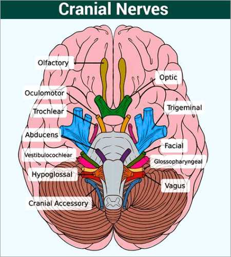
The twelve cranial nerves' names and primary functions are as follows:
Next TopicBrain Tumor Symptoms
|
 For Videos Join Our Youtube Channel: Join Now
Feedback
- Send your Feedback to [email protected]
Help Others, Please Share


