| |

Brain DevelopmentIt is an amazing feat of biological engineering that the human brain can be created from the tip of a 3-millimeter neural tube. The brain develop during pregnancy at a pace of around 250,000 nerve cells per minute to reach the more than 100 billion neurons that make up the typical complement of a newborn. However, the creation of a human brain is astonishing to fathom for reasons other than just the amount of growth. Though the feat of growing a human brain occurs in hundreds of millions of people every year, it is astounding in its complexity, given the vast number of functions that the brain reliably performs and the specificity with which these are assigned to one or another type of cell or small location in the entire assembly. The roughly 100 trillion connections in the brain are the physical foundation for its complexity and speed. However, how is a network this complex first built? Does the fertilized egg's genetic makeup already have a complete set of construction instructions for the human brain, in which each cell is generated as a little piece of the larger design? Moreover, suppose the set of instructions is narrow and precise. How could chance, random mutations, or the impact of the environment have played a part - as they so obviously have done and continue to do - in the formation of the earliest human brains? 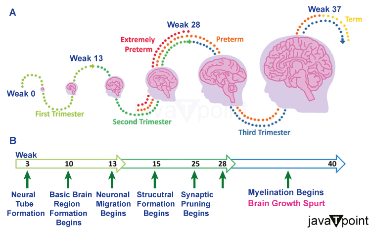
These questions make it clear that any scientific explanation of the evolution of the human brain must overcome a difficult obstacle. Such an account must not only explain a sequence of development with great orderliness and effectiveness but also leave room for the creative effects of chance, such as random mutations and the subsequent natural selection, which have contributed to the spread of this particular type of brain in the first place. Most developmental neuroscientists responding to this question today propose a sequence of phases where the impacts of random external events and built-in instructions are intriguingly mixed. Early Stages of Brain DevelopmentThese stages are:
These events do not happen in a strict order; instead, they overlap in time. When a child reaches the age of around 18 months, no new neurons are created, and the division of different cell types into separate regions is essentially complete. However, eliminating unnecessary connections, which is crucial for determining how the adult brain develops, goes on for years. Numerous productive avenues of neuroscience research have been developed thanks to this concept of brain evolution. Regarding the point at which development was disrupted, it may, among other things, explain well-known brain or nervous system birth abnormalities. Anencephaly, or "without brain," is a disorder that usually causes stillbirth or short-term survival if the neural tube fails to seal correctly very early. In this case, the cells that should create the forebrain and the skull and scalp surrounding it may not be produced. Spina bifida, or a "split spine," is where the spinal column lacks the bone covering of part of its vertebrae. Less severe neural tube anomalies may cause varied degrees of this condition. X-ray exposure, heavy drinking, certain medicines, and a mother's infection with certain illnesses like rubella (German measles) may all adversely affect a child's development during critical early stages. By the seventh month, the fetus starts sending its brain waves, which can be detected via the mother's belly, and the number of brain cells in the cerebral cortex has increased significantly between 15 and 20 weeks following conception or approximately the halfway point of pregnancy. Even though it is still essential up to birth and for some time later, many lines of evidence indicate that appropriate nutrition is most vital for brain development at this point. However, the developing brain has a remarkable potential to rebound and continue its normal development even when subjected to environmental insults like starvation, as long as the adverse conditions are removed during the first three months of life. One of their challenges is that neuropathologists who study congenital neurological abnormalities often investigate the brain years after the aberrant events have occurred. The "normal" data in this field may have a considerable range of fluctuation, which is another issue. A fundamental knowledge of normal brain development is necessary to identify and treat those circumstances that may prevent the development of a healthy brain. Only by initially looking at the prenatal months can we fully understand how the brain develops throughout the formative years of infancy. The long-term process of brain development starts around two weeks after conception and lasts until early adulthood, roughly 20 years later. While there is no doubt that the environment can impact brain development during the prenatal months, it is well known that both the absence of nutrition (such as folic acid) and the presence of toxins (such as alcohol) can have negative effects. On the other hand, postnatal brain development is heavily influenced by experience and the interplay between genes and the environment. The structural alterations that define the first phases of brain development are briefly described here. 1. NeurulationThe growing embryo is organized into a three-layered, spherical form around two weeks after conception. The cells in one region of this sphere thicken to create the neural plate. Then, like a zip, this plate folds over, creating a tube that progressively shuts at the bottom and the top. This results in the development of the neural tube, whose outside cells will give birth to the autonomic nervous system (nerves outside the brain and spinal cord) and whose inner cells will establish the central nervous system (brain and spinal cord). The closed neural tube develops into a three-vesicle structure and quickly transforms into a five-vesicle structure. The several tissue clusters surrounding the ventricles will develop into separate brain structures. The forebrain, which contains the cerebral hemispheres, the diencephalon (the thalamus and the hypothalamus), and the basal ganglia, will develop in the front part of the tube. The midbrain, which connects the diencephalon to the hindbrain, will develop from the cells surrounding the middle vesicle. The hindbrain, which includes the medulla oblongata, the pons, and the cerebellum, will develop from the rear-most part of the tube. Finally, the spinal cord will develop from the cells that are left. 2. ProliferationThe cells that line the deepest portion of the neural tube, known as the ventricular zone, multiply at a logarithmic rate after the overall structure of the tube has been established. A second zone, known as the marginal zone, which will include axons and dendrites, is formed as these cells grow. Since this proliferative period lasts for a while, the newborn brain will have a much higher number of neurons than the adult brain. Apoptosis, or programmed cell death, finally balances off the overproduction of neurons. Apoptosis, which is entirely governed by genetics, causes a reduction in cell counts to adult levels. 3. Cell MigrationThe cells go to their ultimate locations after being born. The stratified tissue that makes up the cerebral cortex is several millimeters thick. It is created by the inside-out migration of cells, starting in the ventricular zone and moving through the intermediate zone before arriving at their ultimate location outside the growing brain. The first migrating cells occupy the deepest cortical layer, whereas the successive migrations go through already-formed layers to create the outer layers. The brain will have generated all six layers around 25 weeks after conception. About 70%-80% of migrating neurons, the majority of which are pyramidal neurons and glia, follow the inside-out pattern of migration described here, known as radial migration. The big neurons in the cortex, known as pyramidal neurons, are in charge of transmitting signals to various layers of the cortex as well as to other regions of the brain. Nonneuronal brain cells called glia help neuronal functions by creating myelin or clearing away waste materials like dead brain cells. Interneurons, comparatively smaller neurons engaged in communication between pyramidal cells in a specific cortex layer, migrate tangentially in contrast. 4. DifferentiationA neuron typically takes one of two routes after reaching its destination: either it differentiates into a mature neuron with axons and dendrites, or it undergoes apoptosis, which causes it to retract. According to current estimations, between 40% and 60% of neurons are thought to retract (Oppenheim & Johnson, 2003). Growth cones, which are minute structures that arise at an axon's border, aid in the development of axons. The cellular activities near the growth cone encourage development in one direction and away from another. In addition to anatomical features near the development cone's tip, such processes are influenced by molecular guidance signals. Aizawa et al. (2004) suggested that the genes governing calcium-regulated transcription factors are the source of a somewhat different mechanism that is assumed to be responsible for dendrite development. When they first emerge from the cell body, early dendrites resemble thick strands with few spines (small protuberances). It becomes more likely for a dendrite to come into touch with an adjacent axon as dendrites grow because of an increase in the number and density of spines. As discussed below, synaptic connections between neurons are the building blocks for brain activity. These connections between dendrites and axons constitute the foundation for these connections. 5. SynaptogenesisA synapse is a junction where two brain cells, typically two neurons and commonly a dendrite and an axon, come into touch. Generally speaking, the first synapses are seen by the 23rd week of pregnancy (Molliver et al., 1973), albeit the production peak does not happen until sometime in the first year of life. Synapses undergo a huge overproduction followed by a slow decline, much like neurons. A significant portion of learning in the early years of life is based on this process of synapse reduction, or pruning, which is extremely experience-dependent. The peak synapse creation occurs at different times in the different brain areas, an essential aspect to remember. Somewhere between the fourth and eighth postnatal month is when the visual cortex, for instance, reaches its apex, but the prefrontal cortex only reaches the fifteenth postnatal month. The time of peak synapse creation varies, and this timing variation is significant because it influences how quickly these areas become plastic; the later the peak synapse production, the longer the region is plastic. 6. Synapses PruningFollowing an excessive synaptic overproduction, the excess synapses are cut down. Up to synaptogenesis, most of the phases of brain development are determined by genes. The pruning process is mostly experience-driven as the brain reaches the stage when connections are removed; however, the balance changes. The timing of synapse pruning is also influenced by the brain region in which it takes place, much like synapse creation. Pruning is finished between the fourth and sixth year of life, for instance, in the regions of the brain responsible for visual and aural perception. In contrast, during adolescence, pruning in regions connected to higher cognitive skills, such as inhibitory control and emotion regulation, persists (Huttenlocher & Dabholkar, 1997). The processes of excessive synaptic growth followed by synaptic shrinkage are crucial to the growing mind's flexibility and capacity for adaptation. It enables the person to react to the particular surroundings in which he or she was born. Environment-activated pathways are reinforced, while inactive pathways are shut off. This allows for adjusting and modifying the neural networks crucial for forming behavior. 7. MyelinationMyelination is the name given to the last procedure in the brain's development. As a result of this insulation, myelinated axons can conduct electrical impulses more quickly than unmyelinated axons, which eventually promote neuronal activity and communication. Depending on the area of the brain it happens in, myelination progresses at different times. In a process that is finished around the preschool years, some sensory and motor regions of the brain get myelinated early. However, the process is finished in adolescence or early adulthood in parts of the brain that are engaged in higher cognitive functions, such as the prefrontal cortex. SummaryBrain development generally starts a few weeks after pregnancy and is believed to be finished by early adulthood. The prenatal and early infancy years are when the brain's fundamental structure is predominantly established, and throughout time, neural networks continue to evolve and become better. The several functions of the brain do not all develop at the same time or along the same timeline. Although the fundamental sense and perception systems are completely formed when kids are in kindergarten, the memory, decision-making, and emotional systems continue to grow throughout childhood. However, many of these skills are built from the ground up in the formative years.
Next TopicBrain Facts
|
 For Videos Join Our Youtube Channel: Join Now
Feedback
- Send your Feedback to [email protected]
Help Others, Please Share


