| |

CytoskeletonCytoskeleton refers to the cytoplasmic structures that provide mechanical support, shape and strength to the cell just like a human skeletal provides to the human body. These structures are made of proteins and can be very minute, fibrous, filamentous and tubular. We can say that cytoskeleton is a network of protein fibres in the cytoplasm, which acts as a structural framework for the cell and thus provides stability to the cell as well as helps in cell movement. It is made of different types of proteins such as tubulin, actin, myosin, tropomyosin, keratin, and more. The cytoskeleton is a unique feature of eukaryotic cells as it is present only in eukaryotic cells and absent in prokaryotic cells such as in bacteria. It is a flexible or dynamic structure as it does not have any fixed shape or its shape may change under different conditions or environments. The various internal cellular structures like the nucleus and cell organelles are able to hold their positions inside the cell and maintain a safe distance between each other due to the cytoskeleton. The major components of the cytoskeleton are described below: 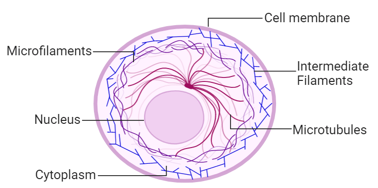
i) Microtubules: They are small hollow tubes like structures made of a single type of globular protein, called tubulin. They are found in the matrix of eukaryotic cells. The structures that help in the movement like cilia and flagella are also made of microtubules. They are also found in the basal granules, aster rays, and axoneme of the sperm tail. The centriole and spindle fibres that take part in the cell division during the metaphase and anaphase state of cell division are also made of the microtubule. So, they play a major role in cell division. The microtubule has a 9+0 arrangement in the centriole. The wall of the microtubule is made of the globular protein tubulin. Tubulin protein is a dimeric as it is made of two subunits alpha-tubulin and beta-tubulin proteins. These subunits have the same three-dimensional structure, which allows them to fit tightly. These dimers are arranged in longitudinal rows which are called protofilaments (singular; protofilaments). The wall of the microtubule is made of 13 protofilaments. Tubulin proteins are non-contractile in nature and are spirally arranged to form the wall of the microtubules. The protofilament is asymmetric as it has alpha-tubulin at one end and beta-tubulin at the other end. The average diameter of the microtubule is 25 nm. Where the diameter of the lumen is around 15 nm. So, the thickness of the wall is around 5 nm. A microtubule can be so large that it may extend across the length and breadth of the cell. One end of the microtubule is known as the plus end and it is terminated by beta-tubulin subunit. The other end is known as the minus end, which is terminated by alpha-tubulin subunits. This polarity is very beneficial for its growth and enables it to participate in mechanical activities. During the growth of the microtubule, the individual dimers (alpha and beta subunit) bind to the plus end and some dimers are released from the minus end. This process continues that helps maintain the length of the microtubule. Functions of microtubules:
ii) Intermediate filaments: They are made up of different types of proteins such as keratin, desmin, nestin and vimentin. Their length is around 10 nm, which is more than the length of the microtubule (25 nm), but less than the length of the microfilament (7 nm). So, they are called intermediate filaments. They are very stable and provide shape and mechanical strength to the nuclear membrane and anchor cell organelles. Unlike other components of the cytoskeleton, they do not take part in cell motility. Functions of intermediate filaments:
iii) Microfilaments: They are solid, unbranched, rod-like structures, so they do not have lumen inside them. They are found in eukaryotic cells and also known as actin filaments. They are very thin as compared to other parts of the cytoskeleton. Microfilaments are made up of contractile proteins which include actin and myosin proteins which are strong as well as flexible and thus very useful in cell movement. Actin is a globular protein, whereas, myosin is a filamentous protein. Functions of microfilaments:
Next TopicCell Membrane
|
 For Videos Join Our Youtube Channel: Join Now
Feedback
- Send your Feedback to [email protected]
Help Others, Please Share


