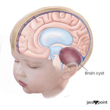
Brain Cyst
A brain cyst or cystic brain lesion is a fluid-filled sac in the brain. They could be benign (cancer-free) or malignant (cancer-causing). Benign refers to the growth not spreading to other body regions. A cyst may have additional materials, such as pus or blood. Cerebrospinal Fluid (CSF) can occasionally be found in cysts in the brain. The brain and spinal cord are covered and cushioned by CSF, a transparent liquid. Some brain tumours develop prior to birth.
A brain cyst can still be problematic even if it isn't malignancy. Headache, vision issues, or nausea are just a few of the symptoms that the cyst may produce by pressing against brain tissue. If this occurs, you might require surgery to have the cyst removed. When a cyst is tiny, stable, and unlikely to develop symptoms, your doctor may suggest observing it rather than doing surgery.
Different Types of Brain Cysts
- Leptomeningeal Cyst (Arachnoid Cyst): This cyst is located between the arachnoid membrane and the brain. One of the protective coverings that surrounds the brain is this membrane. CSF is present in an arachnoid cyst. Although they can occur in adults, these are more frequently seen in children. Males than females are more likely to get this form of cyst.
- A Globular Cyst: This cyst is packed with gel. It frequently develops in one of the brain's four ventricles. The brain's CSF reserves are the ventricles. The third ventricle is frequently the site of colloidal cysts. This is located in the centre of the brain. The cysts may induce intermittent CSF flow obstruction and positional headaches. These are headaches that develop as a result of specific body positions. These typically start to show up as adults.
- A Cystic Dermoid: This cyst is uncommon. It develops when a few epidermal cells become stuck during the prenatal development of the brain and spinal cord. Even sweat gland or hair follicle cells have been seen to be found in these cysts. These frequently show up in kids.
- Cyst on the Epidermis: A different name for this is an epidermoid tumour. It develops when some tissue becomes imprisoned during the formation of the brain and spinal cord, much like a dermoid cyst does. There are no sweat glands or hair follicle cells in epidermoid cysts. They develop slowly. These cysts frequently develop in adults for the first time.
- A Pineal Cyst: On the pineal gland at the centre of the brain, this takes place. Often, imaging examinations performed for another purpose are the only times this sort of cyst is detected. Pineal cysts rarely result in issues. If they do become large, they may occasionally impair vision. Any age group can develop them.
- Brain Infection: Anywhere in the brain, either as a single cyst or several cysts, this can occur. A bacterial infection is frequently the cause of abscesses. They can occasionally be brought on by a parasite or fungus.
- A Cystic Tumour: A benign or malignant tumour is to blame for this. Metastatic refers to a brain tumour that originates external to the brain.
What Causes a Brain Cyst?
The accumulation of fluid in the brain results in brain cysts. During the first few weeks of a baby's development in the uterus, brain cysts can develop. Some cysts may develop as a result of a brain trauma, such as a head injury. In some situations, there might be a connection between a brain cyst and either a cancerous or noncancerous tumour.
What Signs Indicate a Brain Cyst?
The symptoms are typically based on the region of the brain where the cyst is developing. A tiny cyst could occasionally go unnoticed. Before they become huge, certain cysts are "silent" (don't create any symptoms). In some circumstances, you can have an issue with the region of the brain where the cyst is developing. In other situations, a stoppage in the normal CSF flow may be the cause of the symptoms. The intracranial pressure (ICP) in the brain may rise as a result.
Although individual symptoms may vary, they may include:
- Headache (common)
- Face discomfort
- Seizures (rare)
- Nausea and vomiting
- Vertigo or disorientation
- Hearing or vision issues
- Trouble balancing and walking
How are Brain Cysts Identified?
Doctor might find a brain cyst in some circumstances when it appears on an imaging scan that was conducted for another reason. In other situations, you can be experiencing cyst-related symptoms. Your primary healthcare provider has the authority to recommend a neurologist to you. This healthcare provider specialises in diagnosing and managing central nervous system disorders. Or perhaps a neurosurgeon will be suggested to you. This doctor does spinal cord or brain surgery.
A physical examination and a health history are the first steps in the cyst diagnosis process. Your doctor will inquire about your symptoms and previous medical issues. The history of your family's health may also be brought up. Nervous system testing may be part of the physical examination. To examine the brain, imaging techniques could be used. To make the photos show more detail, contrast dye may be applied. The examinations could involves-
- CT Scan: This imaging technique creates precise images of the body using X-rays and a computer. Your spinal cord and brain may be scanned.
- MRI: Photographs of the body are created during this examination using a computer and big magnets. MRIs of your brain and spinal cord may be performed in order to understand more about the cyst and the tissues around.
Repeated scans can be used to determine whether the cyst is expanding.
How are Brain Cysts Handled?
Your doctor might suggest having a brain cyst surgically removed if it's causing issues. Your doctor may decide to perform frequent brain scans to keep an eye on the cyst if it is not spreading or producing symptoms. The type of cyst can affect the course of treatment. As an illustration: If you have an arachnoid cyst, your doctor might pierce the cyst sac and drain the fluid. The fluid is either removed with a needle or drained into the CSF. The sac may re-fill with fluid if your healthcare professional drains the cyst without removing a portion of it or inserting a permanent draining tube.
- Your doctor will probably remove any dermoid or epidermoid cysts you have. Most likely, the entire cyst and its sac will be removed.
- Colloid cysts frequently result in a buildup of extra CSF, which may return if the cyst is not completely removed. We call this hydrocephalus. This may result in a risky rise in intracranial pressure.
- Some of this pressure can be released via a shunt or drainage tube. Because they are frequently found deep into the brain, removing colloid cysts can be challenging.
- Your healthcare provider may employ specialised surgical techniques to remove them, which may involve inserting tiny endoscopic tools through a thin tube into the brain. These may frequently be controlled by keeping an eye out for any changes.
- Chemotherapy, radiation, or surgery are all options for treating tumour cysts. Both of these options are possible.
- Antibiotics, antifungals, and antiparasite medications are used to treat abscesses. Surgery can also be required.
Key Points of a Brain Cyst
- Fluid-filled sacs called brain cysts can develop in the brain. They may be benign (not cancer) or malignant (cause cancer).
- Both toddlers and adults can develop brain cysts. Brain cysts can sometimes start before birth but not show symptoms until much later.
- On occasion, a brain cyst that is asymptomatic will be detected during an imaging scan performed for another reason.
- Brain cysts can be of several forms. Arachnoid, colloid, dermoid, epidermoid, pineal, infectious, and tumour cysts are among those that fall into this category.
- Even if a brain cyst isn't cancerous, it might press against brain tissue and create symptoms. A cyst may occasionally prevent the normal flow of CSF. This may lead to issues.
- Your neurosurgeon or neurologist might suggest removing the cyst surgically. Instead of watching the cyst to see if it remains stable, your healthcare professional can advise doing so if it is small and unlikely to produce symptoms.
Next Step
Here are some pointers to help you make the most of a visit to your doctor:
- Be aware of your visit's purpose and your goals.
- Make a list of the questions you want answered before your visit.
- Bring a companion to assist you remember the information your provider gives you and to ask questions.
- Take note of any new diagnoses, medications, treatments, or tests during the appointment. Additionally, make a note of any fresh directions your provider offers you.
- Understand the benefits of any new medications or treatments that have been prescribed for you. Also be aware of any potential negative effects.
- Enquire about alternative treatments for your condition.
- Recognise the rationale behind a test or procedure recommendation and what the results may mean.
- Know what to expect if you forego the examination, operation, or medicine.
- If you have a follow-up appointment, write down the date, time, and purpose of the visit.
- Keep your provider's contact details handy in case you have questions.
|

 For Videos Join Our Youtube Channel: Join Now


