| |

Brain WavesThe central nervous system's rhythmic or recurring patterns of neural activity are known as brainwaves or neural oscillations. Numerous processes, including those found in individual neurons and connections between neurons, can generate oscillatory activity in neural tissue. Oscillations may manifest in a single neuron as changes in membrane potential or as periodic patterns of action potentials, which cause postsynaptic neurons to oscillate. An electroencephalogram may show macroscopic oscillations at the level of neuronal ensembles caused by the synchronized firing of many neurons. In most cases, feedback connections between neurons cause the synchronization of their firing patterns, which leads to oscillatory activity in groups of neurons. Different frequencies from the firing frequency of individual neurons might result from oscillations caused by neuronal contact. The macroscopic brain oscillations known as alpha activity are a well-known example. Researchers began noticing human neural oscillations as early as 1924 (by Hans Berger). Intrinsic oscillatory behavior was discovered in vertebrate neurons more than 50 years later, although its functional significance is still not entirely understood. The creation of rhythmic motor output, feature binding, and information transmission pathways are some of the potential functions of brain oscillations. More knowledge has been obtained in recent years, particularly due to improvements in brain imaging. Understanding how oscillations are produced and what functions they serve is an important topic of study in neuroscience. At various organizational levels, oscillatory activity in the brain is often seen, and it is believed to be crucial for processing neural data. A physiological relevance for brain oscillations is supported by several experimental research, although a consistent explanation is still missing. HistoryIn 1875, Richard Caton published his research on the electrical activity he had found in the brain hemispheres of rabbits and monkeys. Adolf Beck reported his findings on the electrical activity that occurred in the brains of dogs and rabbits in 1890. These findings included rhythmic oscillations that were affected by light and picked up by electrodes that were put right on the brain's surface. Vladimir Vladimirovich Pravdich-Neminsky reported the first animal EEG and the dog's evoked potential before Hans Berger. Types of Brain WavesAs already discussed, several states are related to various brain waves. The frequency of brain waves is measured in Hertz (Hz) cycles per second and varies greatly. When slower brain waves predominate, we may feel lazy, disorganized, and distracted. We may also experience depression or sleep problems. Higher frequencies may cause critical thinking, hypervigilance, anxiety, nightmares, and impulsive conduct. They can also cause critical thinking, hypervigilance, and worry. 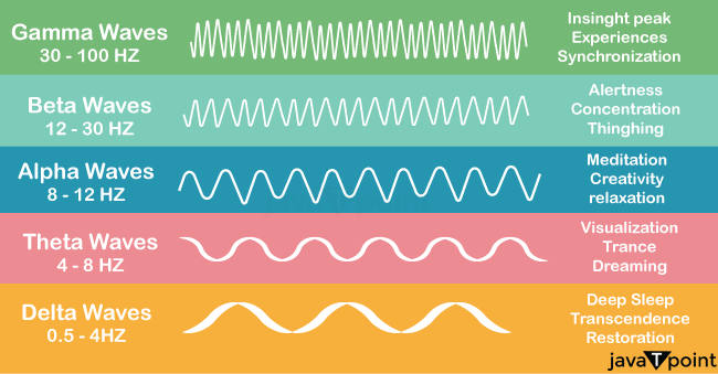
1. Delta WavesSlow brainwaves called delta waves (1-4 Hz) start in stage 3 of the sleep cycle and take over practically all EEG activity by stage 4. This stage is considered crucial for the therapeutic effects of sleep because it stimulates repair and regeneration. Concentrating when a person has too many delta waves when awake may be very challenging, which can cause learning problems and ADHD. It has been shown that people with different kinds of brain lesions create delta waves when awake, which makes it very challenging to carry out cognitive activities. Sleep, walking, and talking happen when delta output is strong. According to research, higher-frequency delta rhythms are produced by thalamocortical cells and intracortical network connections, while cortical circuits only produce delta rhythms below 1 Hz. Importantly, delta could also represent the activity of acetylcholine and dopamine. Delta is strongly associated with the physiological interface between the brain and the body because it is active within brain networks that link the cortex and insula with the hypothalamus and the brainstem. Gamma-aminobutyric acid (GABA) universally inhibits neurons during delta-wave sleep. 2. Theta WavesDaydreaming and sleep heavily involve theta waves (4-8 Hz). Young children commonly experience cortical theta, whereas older kids and adults typically experience it during contemplative, sleepy, or sleeping states (although not during the deepest sleep phases). A typical symptom of ADHD is excessive theta activity, which may make us feel disorganized or daydream while awake. It is believed that excessive theta in the left hemisphere leads to disorganization, while excessive theta in the right hemisphere leads to impulsivity. People with attention difficulties often have higher theta activity in the brain's frontal lobes. Frontal Midline Theta: An ERP is typically sinusoidal and strong in amplitude (1-10 second bursts) and happens in reaction to stimuli. The hippocampus's sensory gate is known to open during this midline theta, allowing for the intermediate storage of episodic information. Frontal midline theta has a frequency range of 5-7.5 Hz, with an average of 6 Hz. This rhythm is linked to episodic encoding, retrieval, and working memory. Deep concentration and hypnosis are other times when it manifests. The anterior cingulate is assumed to be the source of the frontal midline theta. It mostly manifests during concentrated concentration-intensive tasks, and as the workload grows, so does its amplitude. It is mostly centered in the area of Fz. The signal is diminished or perhaps completely removed when tense and restless. The signal is recovered after taking medication for anxiety. This shows that the anterior cingulate cortex controls emotions, from agitated worry to concentrated calm. Hippocampal Theta: has been discovered in the amygdala, entorhinal cortex, hypothalamus, and posterior cingulate. Often more diffuse and tonic, it stimulates and synchronizes memory. 3. Alpha WavesAlpha waves (8-12 Hz) predominate during peaceful contemplation periods and other meditation states. Being now and in the current moment is what is referred to as the "power of now" or alpha. Like an automobile idling at a stoplight, it is the brain's resting state. Overall, mental coordination, relaxation and attentiveness, mind-body connection, and learning are all aided by alpha waves. Right hemisphere alpha levels tend to be higher, while low alpha levels are linked to undesirable habits like social retreat. Additionally, this is seen in depressed individuals, particularly in those with excess frontal alpha. The active and sufficient suppression of the unnecessary sensory pathways is mediated by alpha. The resonance process between the thalamus and the cortex results in the production of alpha, which is connected to resource allocation in the cortex. Alpha may be viewed as the method by which the sensory gate to the cortex can be closed if we consider the thalamus to be its entrance. In addition to participating in binding processes and resource allocation about orientation and task sequences, alpha is tightly associated with reticular activation. Alpha declines when sleep begins while concentrating on activities, and is also a typical effect of aging. It is often a sign of pathologically slowed high-amplitude alpha, linked to Parkinson's disease and cognitive loss when alpha slows, and theta rises in frequency. This shows deterioration of myelination, cell death, and increasing metabolic inefficiency in the brain. The well-functioning brain exhibits elevated levels of alpha after finishing a task and receiving feedback. This is related to post-reinforcement synchronization, often known as PRS, consolidating task events. This corresponds to an alpha burst in the brain while information is consolidated. 4. Beta WavesWhen our attention is focused on cognitive processes and the outside world, beta waves (12-38 Hz) reflect our typical waking state of awareness. When we are aware, attentive, and engaged in problem-solving, decision-making, and focused mental work, Beta is our brains' dominant "fast wave" activity. Strong Beta (22-38 Hz) is supposed to be extremely complicated thinking, integrating new experiences, strong anxiety, or excitement. Low Beta (12-15 Hz) is regarded as "fast idle" or pondering thought. Beta (15-22 Hz) indicates high engagement and actively figuring things out. Continuous high-frequency processing is ineffective for our brains to function; it may cause stress and make it difficult to unwind. If it occurs at night, it can also make settling the mind and falling asleep difficult. Beta waves predominate more in the left hemisphere, while too much Beta in the right hemisphere has been linked to manic symptoms. There are differences in how the brain's three beta and gamma levels allocate their space. Higher beta frequencies are generally believed to be more linked with arousal. However, others argue that they are mostly the product of muscular artifacts. For instance, Helleter et al. discovered that anxiety was strongly connected with heightened right hemisphere beta. At the same time, more recent research has shown that headaches and sleeplessness correlate with greater temporal lobe beta frequencies. 5. Gamma WavesThe highest-frequency brainwaves are gamma waves, which oscillate between 30 (about) and 100 Hz. They are linked to high levels of cognitive performance and peak focus. Low levels of gamma activity have been associated with cognitive impairment, poor mental processing, and poor memory. In contrast, high levels of gamma activity are associated with intelligence, compassion, good memory, and happiness. Gamma is now of little therapeutic use because muscle contamination prevents its accurate measurement using current EEG equipment, according to the argument. Despite encouraging studies suggesting that Gamma training may improve intellect, it will be of meaningful therapeutic use once this technological problem is overcome. Gamma and theta attract neurons, which promote activity in the nearby cell column. As a result, it is connected to cortical processing linked to cognitive activities and may be connected to meditative states. Still, the evidence for this connection needs to be more extensive. Brain Waves and ECGAll levels of the central nervous system exhibit neural oscillations, which may be detected by electroencephalography (EEG), including spike trains, local field potentials, and large-scale oscillations. Oscillations may often be identified by their frequency, amplitude, and phase. Time-frequency analysis may be used to derive these signal features from brain recordings. Amplitude variations in large-scale oscillations are thought to be the consequence of modifications to the local synchronization of a neuronal ensemble. The oscillatory activity of distant brain structures (single neurons or neuronal ensembles) may synchronize in addition to local oscillatory activity. Numerous cognitive processes, including information transmission, perception, motor control, and memory, have been associated with neural oscillations and synchronization. Neural isolation occurs when the electrical activity of neurons is not temporally synchronized, in contrast to neuron synchronization. The possibility that the neuron will hit its threshold potential and let the signal pass to the next cell reduces at this point. This phenomenon is often seen when the spectral intensity falls from the total of these neurons firing, and it may be used to distinguish between neuronal isolation and cognitive activity. The difficulty of the signal to spread to neighboring neurons, an indication of deterioration (such as hypoxia), has been characterized using novel non-linear approaches that combine temporal and spectral entropic correlations concurrently. The brain activity produced by massive clusters of neurons has been the subject of the most extensive study of neural oscillations. Techniques like EEG may be used to quantify intense activity. Pink noise-like spectrums are typical of EEG signals but also show rhythmic activity in some frequency regions. Alpha activity (8-12 Hz), which may be picked up from the occipital lobe during calm wakefulness and rises while the eyes are closed, is the earliest and best-known frequency band. Theta (4-8 Hz), beta (13-30 Hz), low gamma (30-70 Hz), and high gamma (70-150 Hz) are other frequency bands. Faster rhythms like gamma activity have been connected to cognitive processing. In fact, EEG signals fluctuate significantly during sleep and demonstrate a shift from faster to slower frequencies, such as alpha waves. The spectral content of sleep at various phases is often used to define them. Consequently, cognitive states like awareness and consciousness have been associated with neuronal oscillations. Although EEG recordings are the primary method used to study neural oscillations in human brain activity, other, more intrusive recording methods, such as single-unit recordings, have also been used to monitor them. Neurons in rhythmic patterns may produce action potentials or spike patterns. Resonators, or certain kinds of neurons, fire at specific frequencies. Another kind of rhythmic spiking is bursting. Spiking patterns are seen as essential for the coding of information in the brain. In the absence of action potentials, oscillatory activity may also be seen as subthreshold membrane potential oscillations. Local field potential oscillations may result from several neurons firing at once. In recorded data, the strength of brain oscillations may be estimated using quantitative models. In "neurodynamics," a branch of cognitive science that emphasizes the dynamic aspect of neural activity in defining brain function, neural oscillations are often examined within a mathematical framework. It employs differential equations to explain how neural activity changes over time and treats the brain as a dynamical system. It specifically tries to link dynamic brain activity patterns to mental processes like perception and memory. Neural oscillations may be analytically analyzed in a fairly abstract fashion. Computer simulations of a computational model are often used to study oscillatory activity in a more physiologically accurate environment. Brain oscillations have many diverse uses, which change depending on the kind of oscillatory activity. Examples include the neuronal binding of sensory elements in perception, such as the form and color of an item, and the creation of rhythmic activity, such as a heartbeat. Numerous neurological illnesses, such as excessive synchronization during seizure activity in epilepsy or tremor in Parkinson's disease patients, are also impacted by neural oscillations. A brain-computer interface or other external devices may be controlled via oscillatory activity. PhysiologyAll organizational levels of the central nervous system exhibit oscillatory activity. The activity of a single neuron at the micro-scale, a small group of neurons at the mesoscale, and the activity of several brain areas at the macro-scale have all been generally recognized as three distinct levels. MicroscopicNeurons produce action potentials due to variations in the electric membrane potential. Neurons may produce multiple action potentials in succession to create spike trains. The brain's neuronal coding and information transmission are based on these spike trains. Spike trains often exhibit oscillatory activity and may generate rhythmic spiking and bursting patterns. Sub-threshold changes in membrane potential may also be used to detect oscillatory activity in a single neuron. Since these periodic variations in membrane potential do not cross the crucial threshold, no action potential is produced. They may result from inherent characteristics of neurons or postsynaptic potentials from synchronized inputs. The patterns of neuronal spiking may be used to categorize it. Neuronal excitability may be categorized into Class I and Class II. Class I neurons may produce action potentials at arbitrarily low frequencies depending on the input power. Still, Class II neurons produce action potentials in a specific frequency range that is largely insensitive to variations in input strength. Sub-threshold oscillations in membrane potential are another characteristic of class II neurons that are more common. MesoscopicA group of neurons may also produce oscillatory activity. The firing rhythms of several neurons may synchronize via synaptic contacts, and the rhythmic variations in electric potential brought on by their action potentials will add up (constructive interference). In other words, synchronized firing patterns cause synchronized input into other cortical regions, which causes oscillations of the local field potential with enormous amplitudes. Electroencephalography (EEG) and magnetoencephalography (MEG) may be used to measure these big oscillations external to the scalp. Since single neurons produce electric potentials that are much too minute to detect outside of the scalp, EEG or MEG activity always represents the synchronized activity of hundreds of millions of neurons with a comparable spatial orientation. Rarely do all neurons in a neuronal ensemble fire simultaneously or completely synchronized. Instead, neurons are more likely to fire simultaneously due to regularly regulated firing probability, which causes oscillations in their mean activity (see image at the top of page). As a result, the froptionalrge-scale oscillations don't need to coincide with the pattern of firing of individual neurons. However, in the intact brain, cortical cells are often assaulted with rapidly variable synaptic inputs and seem to fire at random. Isolated cortical neurons activate routinely under certain circumstances. However, the likelihood of a large group of neurons will cause oscillations in the mean-field if it is periodically modulated at a common frequency (see also graphic at the top of page). Through regional connections between excitatory and inhibitory neurons, neural ensembles may produce oscillatory activity on their own. By creating a small window for effective stimulation and regularly adjusting the firing rate of excitatory neurons, inhibitory interneurons contribute significantly to creating neuronal ensemble synchronization. MacroscopicAdditionally, interactions between various brain regions connected by the structural connectome might lead to neural oscillation. Delays in time are significant in this case. These connections between brain regions create feedback loops since all brain areas are bidirectionally connected. In oscillatory activity, frequency and delay time negatively correlate in positive feedback loops. The thalamocortical radiations, which link the thalamus and cortex, are an example of such a feedback loop. Recurrent thalamocortical resonance, an oscillating activity, may be produced by this thalamocortical network. In the production of alpha activity, the thalamocortical network is crucial. The partial synchronization of subsets of brain regions oscillating in the gamma band (created at the mesoscopic level) leads to oscillations in the beta frequency range in a whole-brain network model with realistic anatomical connections and propagation delays between brain areas.
Next TopicDeep Brain Stimulation
|
 For Videos Join Our Youtube Channel: Join Now
Feedback
- Send your Feedback to [email protected]
Help Others, Please Share


