| |

Image Processing - A Tool For Medical DiagnosticsIntroductionMedical image processing has become a most powerful tool for detecting and treating various disorders in modern healthcare. Image processing is fundamentally the analysis and manipulation of digitalized images, allowing for the extraction of priceless insights from complicated visual data. Explores the elements and uses of image processing, emphasizing how important it is to the field of medical sciences. How to process imagesBefore we go into the particulars of medical applications, let's define the phrase "image processing."It has two crucial parts "image" and "processing." An image visually depicts an object, scene, or data in this context. The term "processing" refers to handling or utilizing this data.
Identifying and diagnosing diseases:MRI (Magnetic Resonance Imaging): MRI is a strong imaging method that combines radio waves and intense magnetic fields to produce fine-grained images of inside body components. In order to improve the quality of MRI scans and make it simpler to spot abnormalities like tumors, injuries, and neurological problems, image processing is essential. CT (Computed Tomography): X-rays are used in CT scans to provide cross-sectional images of the body. It is possible to reconstruct these images into 3D representations with the aid of image processing, which is helpful for precise diagnosis of ailments like internal injuries, vascular disorders, and bone fractures, as well as for surgical planning. PET-MRI and PET-CT: These hybrid imaging procedures, PET-MRI and PET-CT, combine the benefits of both positron emission tomography and MRI/CT. Image processing can provide precise anatomical and functional information by fusing the PET and CT images. This is especially helpful for cardiac evaluations, neurological issues, and cancer staging. Ultrasounds: Images from ultrasounds are improved using image processing techniques, making it simpler to see organs and check the health of growing features during pregnancy. Diagnostic ultrasounds are frequently employed in obstetrics and gynecology. Surgical Techniques: Procedures inside the body are carried out utilizing tiny cameras and small incisions during laparoscopic or minimally invasive surgery. Image processing is essential to give surgeons good, magnified views of the operating area. This improves accuracy and lessens the procedure's invasiveness, resulting in quicker recovery times and fewer problems.
Artificial limbs and prosthetic devices:Measurement and modeling: 3D scans of a patient's residual limb or the area where a prosthetic device will be connected are processed using image processing techniques. These scans aid in developing precise digital models of the limb, enabling the exact personalization of prosthetic devices. Customization: Using these digital models, engineers, and medical specialists may create prosthetic limbs tailored to match each patient's needs and perform well. Alignment and calibration: Image processing supports the alignment and calibration of prosthetic limbs with the patient's body, providing optimal alignment and function to increase mobility and quality of life. 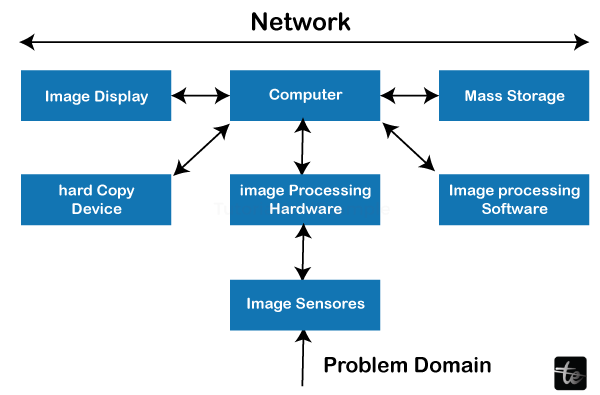
The key steps in this process are as follows:Input:The first step in the cancer diagnosis procedure is to obtain the input data, which frequently consists of medical imaging like MRI scans, CT scans, or X-rays. Identifying probable anomalies or malignant areas within the patient's body depends on these photographs. Preparation:An essential step in getting the input images ready for analysis is pre-processing. During this stage, various operations are carried out to improve the photos' quality and eliminate any extra noise or artifacts. Pre-processing methods that are frequently used include: Noise reduction: Eliminating erratic changes or flaws in the image brought on by equipment constraints or patient movement. Image enhancement: Increasing the image's contrast and clarity to make small details easier to see. Normalization: Modifying the image's intensity levels to maintain consistency between scanners and different images. Pre-processing: Makes sure that the input photos are ready for examination, which makes it simpler for algorithms to find potentially malignant areas. Segmentation:Dividing a pre-processed image into separate regions or segments is known as segmentation. Segmentation is used in cancer detection to separate the regions of interest that may include malignant tissues. This stage entails distinguishing between healthy and diseased tissue based on differences in pixel intensity, texture, or other factors, which might be difficult.
Extracting Features:The next stage is to extract pertinent information from these regions after the image has been segmented to find regions of interest. Features represent the qualities of the split areas through quantitative measurements or characteristics. Features in cancer detection could include: Size: The region's dimensions after segmentation. Shape refers to features like roundness, asymmetry, or irregularity. Patterns: Changes in pixel intensities throughout the area are called texture. Intensity: The average pixel value's intensity, variance, or other statistical measurements. Edge Features: Details regarding the region's borders or edges. These extracted characteristics are essential for separating malignant from non-cancerous tissues. These features are frequently used to train classification models in machine learning methods like support vector machines (SVMs), decision trees, or neural networks. Results:Presenting the findings to the medical specialists is the last step in cancer detection. A representation of the image with spots labeled as potentially malignant areas is frequently included in the report. This can be displayed as a separate map or overlays on the original image. Information regarding the discovered regions' size, location, and properties may also be included in the output.
The ability of the medical professionals to diagnose and a treat cancer has been greatly improved by integrating image processing, machine learning, and artificial intelligence into cancer detection workflows. These technologies eventually enhance patient outcomes and treatment options by enabling earlier detection and more accurate localization of malignant areas. Benefits:Early Disease Detection: Image processing methods allow early detection of illnesses and other irregularities in medical imaging. Making this early diagnosis to start therapy promptly and enhance patient outcomes may be essential. Accuracy: Image processing improves diagnosis accuracy by increasing the quality of medical images and obtaining quantitative data. This lessens the possibility of a wrong diagnosis and guarantees that medical practitioners have access to crisper and more educational photos.
Image processing offers objective, quantitative information about a patient's status. This knowledge benefits from making data-driven therapeutic decisions and monitoring illness development and therapy effectiveness. Enhanced Visualization: Image processing makes anatomical structures and anomalies easier to see. This makes it easier for medical personnel to recognize and comprehend complex illnesses. Distant Consultations: Image processing helps telemedicine by facilitating distant consultations and second opinions. Patients in underserved or remote areas can obtain expert medical advice without having to travel. Medical image processing: This is a useful tool for research and field instruction. It aids in creating novel diagnostic procedures, facilitates the comprehension of disease mechanisms, and offers medical professionals and students instructional resources. Cost-Effective Healthcare: Early detection and accurate diagnosis by image processing can lead to cost savings in the long term. It can aid in avoiding pointless examinations, operations, and hospital stays.
Faster Diagnosis: By speeding up the analysis of medical images, automated image processing algorithms can reduce how long patients must wait for their diagnoses.
Continual Improvements: As technology and AI improve, medical diagnostic image processing techniques get more advanced, resulting in continual
Applications:Diagnostics in medicine use image processing in a wide variety of ways. It aids medical personnel in analyzing and interpreting medical pictures to make precise diagnoses, formulate treatment plans, and track the development of diseases. Here are some examples of particular image processing uses in the area of medical diagnosis: Radiology:The quality of X-ray images can be improved using image processing techniques, making it simpler to spot fractures, tumors, and other abnormalities.
Angiography: Image processing aids in the analysis of angiographic images to determine the health of blood vessels, identify blockages, and prepare for procedures like angioplasty. Ultrasound Imaging:Fetal Imaging: Image processing is performed to create 2D and 3D ultrasound images of fetuses. These images help in prenatal diagnosis and monitoring. Cardiac ultrasound: It assists in determining how well the heart is working and identifying any problems. Dermatology:Dermatologists can analyze skin lesions, moles, and rashes using image processing to determine if they are benign or cancerous. Pathology: Image processing enables pathologists to digitize and analyze tissue samples, enabling remote consultation and more precise diagnosis. Ophthalmology:Retinal imaging uses optical coherence tomography (OCT) and fundus photography to diagnose and track retinal illnesses such as diabetic retinopathy and ages-related macular degeneration. Endoscopy: Endoscopies of the gastrointestinal system can detect polyps, ulcers, and cancers using image processing to analyze the images in real time. Nuclear Medicine: Emission of a single photon Image processing aids in the reconstruction and interpretation of functional images, which are used to research organ function, diagnose cancer, and evaluate treatment response in SPECT and PET scans. Dental X-rays: Cone Beam Computed Tomography (CBCT) Image processing enables 3D visualization of dental structures for use in diagnosis, treatment planning, and orthodontic assessments. Radiology for intervention:Image-Guided Surgery: During surgical procedures, image processing makes real-time navigation possible, ensuring accurate tool placement and reducing harm to healthy tissues. Remote Diagnosis: Image processing allows transmitting and analyzing medical images online, enabling doctors to offer remote consultations and diagnoses, particularly in underserved or rural areas. Machine Learning and AI: Image processing techniques are combined with machine learning and AI algorithms to automate the diagnosis and classification of diseases. For example, tumors in mammograms may be recognized, and based on patient data and medical images, certain ailments can be predicted to have a high risk of developing. Disadvantages:While image processing is a potent tool for medical diagnostics with many benefits, it also has several drawbacks and difficulties. The following are some of the main drawbacks of image processing in the medical industry: Cost: Putting sophisticated image processing systems in place and getting the required hardware and software can be pricey. Smaller healthcare facilities or those located in environments with limited resources may find this expense to be prohibitive. Complexity: Creating and maintaining methods and systems for image processing can be challenging. It frequently calls for specialized computer science and medical imaging knowledge, creating a need for more qualified workers. Data security and privacy: Sensitive patient data might be found in medical photographs. A crucial problem in the medical industry is ensuring the confidentiality and privacy of patient data. Interoperability: Various imaging modalities and medical equipment can output images in various formats. Achieving interoperability and seamless data flow between various platforms can be challenging. Radiation Exposure: Ionizing radiation is used in some imaging procedures, including CT scans and X-rays. Patients may have dangers from repeated exposure, so weighing the advantages of diagnosis against any potential concerns is critical. False Positives and Negatives: It's possible that image processing techniques don't always offer 100% accuracy. Potential misdiagnoses can result from false positives. Processing Time: While automating image processing might expedite diagnosis, some sophisticated algorithms may call for lengthy computations. This can cause treatment choices in urgent situations to be delayed. Algorithm Dependence: The efficiency of image processing is highly dependent on the caliber and sophistication of the employed algorithms. Results can be erroneous as a result of subpar or out-of-date algorithms. Strict regulatory standards, such as those established by the Food and Drug Administration (FDA) in the United States, must be met by medical image processing software and systems. It can be time-consuming and expensive to meet these standards.
Next TopicPlot a line along 2 points in MATLAB
|
 For Videos Join Our Youtube Channel: Join Now
Feedback
- Send your Feedback to [email protected]
Help Others, Please Share


