| |

Eye DefinitionThe eye is the bodily organ that grows and controls a person's ability to see. Each component of the eye's structure has a specific function that contributes to preserving or improving vision. By way of the pupil, light enters the eye, and it is bent and shaped by the lens before moving on to the retina and macula, where it produces images that are then conveyed to the brain using the optic nerve. 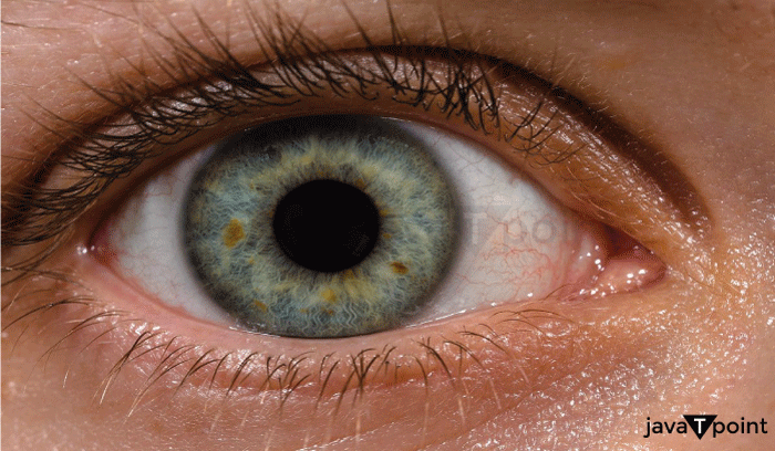
The outer layer of the eye receives light and shields the other layers. The middle layer nurtures the eye and contains most of the blood in the eye. The inner layer receives light and converts it into pictures sent to the brain. A person's eye color and shape might differ significantly from another's. Blue light wavelengths, problems with the eye, and damage all impact the eye's capacity to see. Other animals, like eagles, have different eye anatomy influencing their vision and allowing them to see more clearly or precisely than the human eye. Definition of EyeThe eye is an organ that manages sight and enables vision. It has an asymmetrical globe-shaped find in the head. Various pigmentations or colors can exist in the iris of the eye. The eye is like an organic camera housed in the skull's socket; it can focus on a single object for visual clarity and detail perception while blurring any irrelevant or off-focus elements of the current image. The evolution of the eye still needs to be fully understood. Still, I R Schwab, from the University of California's Department of Ophthalmology & Vision Science, lectured on developing the ocular toolkit. It is believed that the eye evolved from organic mechanics that helped the body maintain a circadian rhythm, which allows us to recognize day and night and know when to rest and be active. The sclera is the term for the eye's white portion. The sclera protects the eye from harm and aids in maintaining the eye's globe-like shape. Human Eye Function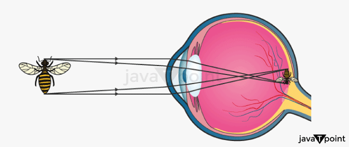
The human eye's pupil receives light, which the lens directs onto the macula and retina. The outer layer of the eye allows the eye to absorb light wavelengths from the environment to produce sight. The cornea, iris, and the white portion of the eye, known as the sclera, are all found in the eye's outer layer. The inner layer of the eye is used by the eye to build a picture from the light that the outer layer of the eye receives. The optic nerve and retina are located in the inner layer of the eye. The choroid and iris are located in the central layer of the eye, which is utilized to nourish the eye. The eye can produce tears because of the three layers found in the tear film. These tears contain oils that offer constant lubrication and nourishment. The eye must be wet to operate effectively. Different Parts of an Eye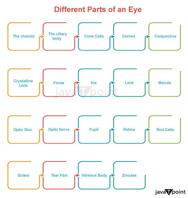
The eye comprises 19 main components listed along with their functions. It provides a detailed understanding of how the eye works. 1. The choroidBetween the sclera and the retina lies a layer of the eye called the choroid. ScienceDirect claims that most of the blood flow in the eyes is accounted for by the choroid, which is mainly made up of blood vessels. The choroid's primary job in the eye is to employ blood flow to feed other crucial eye components, such as the retina, macular, and optic nerve, with nutrition and nourishment. The choroid's second job is to protect the eye from potentially hazardous light and reflection. 'Red eye' in photographs results from the choroid's protecting portion. When the choroid has been harmed, the retina bleeds. Eventually, the deterioration causes both central and peripheral vision to be lost. 2. The Ciliary BodyThe ciliary body, a ring-shaped structure, is attached to the iris. The eye's middle layer consists of three components, one of which is the ciliary body. The choroid and the iris are the other components of the middle layer. The ciliary muscle is responsible for holding the lens in place in the eye while also helping it to change shape and focus, which is part of the role of the ciliary body. The ciliary processes are used by the ciliary body's second purpose, which is to cause the eye's aqueous humor to be produced. The fluid, known as aqueous, transparent, and clear humor, is essential for feeding the eye and maintaining its structure. When the ciliary body is damaged, vision gets blurry, and focusing becomes challenging. Vision will be lost entirely if the ciliary body is damaged and not cared for. 3. Cone CellsCone cells, located close to the eye's macula, aid in our understanding of colors and fine details. Are you aware that there are 6 million cone cells in your eye? These cells are photoreceptors that convert light into signals sent to the brain. Cone cells are one of two types of photoreceptors in the eye. The variants of eye rods are the other sort of photoreceptor. Cone cells come in three varieties: red-sensing, blue-sensing, and green-sensing cones. The purpose of each type of cone cell is to aid the eye in understanding the pigmentation of the cone cells that sense color. Within the eye, cone cells are in charge of color vision. Cone and rod cells' secondary function is to transform light into electric impulses and send them from the optic nerve to the brain. While cone cells are damaged, it makes people more sensitive to light, provides them with less information while looking directly ahead, and makes it more difficult to detect color. 4. CorneaThe National Eye Institute defines it as the transparent outer layer's front portion of the eye. The pupil, iris, and anterior chamber are all immediately covered by it, situated in the very front of the eye. The cornea's primary job is concentrating the received light and regulating how much light gets into the eyes. The amount of light entering the eye affects it; the cornea changes size, becoming smaller or more significant, and bends and refracts the light the eye receives. The cornea, which concentrates most of the eye's light, is the initial point of light contact with the human eye. The second purpose of the cornea is to shield the eye from harmful glare, such as UV radiation from the sun. The cornea can repair cuts, scars, and other minor abrasions. The effects of severe corneal injury are vision distortion and a reduced capacity to see light. 5. ConjunctivaThe conjunctiva is a thin tissue that lines the eyelids and covers the eyes' whites. It looks like a mucous membrane. The tissue covering the conjunctiva is transparent and clear. It is found in the eye's outer layer. The conjunctiva's primary job is to release fluids that keep the eyes moist and shield them from infections, unwanted substances like dust and other bodies, and germs. The conjunctiva comprises three parts: the bulbar conjunctiva, the palpebral conjunctiva, and the fornix conjunctiva. While the bulbar conjunctiva protects the sclera, it does not protect the cornea. The area covering the eyelid's top and bottom sections is the palpebral conjunctiva. The fornix conjunctiva is located between the other two components, allowing the eyelid and eyeball to move freely. The conjunctiva's second job is to prevent unwanted materials like contact lenses from entering the eye from behind. The eyes' appearance is affected by conjunctival damage, resulting in problems including dry eye and impaired vision, discoloration, and apparent eye injury. 6. Crystalline LensThe crystalline lens, a natural focusing lens in the eyes, provides a part of the optical power contained within the eye. It is also sometimes referred to as the eye's lens. The lens's primary purpose is to bend and concentrate light by changing form due to its essential flexibility. The lens directs the light it receives into the retina by utilizing its shifting shape, which converts the outside light into a clear picture. When focusing on distant objects, the lens collaborates with the ciliary muscle. It is its second purpose. The force of the ciliary muscles contributes to the crystalline lens' capacity to alter form. The picture is received upside down when transmitted by the crystalline lens. The optic nerve turns the picture around so that it is right-side up as the light from the crystalline lens travels to the brain. Cataracts, a damaged lens, and impaired vision result from damage to the crystalline lens. Examples of damage that cannot be repaired include scratching or dislocation of the crystalline lens. Because the iris loses its support within the eye's structure when the crystalline lens is damaged, the iris may also be impacted. 7. FoveaIt, also known as the fovea centralis, is a tiny depression or pit in the retina's core that regulates how sharply people can see in the center of their vision. Cone cells that support vision can be found in the fovea centralis and the nearby parafoveal and perifoveal areas. Since the fovea has many cone cells, it serves as the primary structure for central vision clarity and the ability to recognize fine detail. Damage to the fovea impairs one's capacity to see color, the daylight and makes it harder for the eye to focus on fine details. Eye disorders such as macular degeneration can harm the fovea. 8. IrisThe iris, a visible and colorful eye part, is located behind the cornea and in front of the crystalline lens. The iris' primary job is to regulate the pupil's size and the eye's overall light sensitivity. The muscle known as the iris causes the pupil to narrow in reaction to excessive light and enlarge in response to inadequate light. The iris and ciliary body are connected. Based on heredity and the quantity of melanin present in the iris' front layers, the pigmentation in the iris identifies a person's eye color. When the iris is damaged, the eye's appearance and ability to accept light are affected. As a result, there is visual loss and extreme light sensitivity. 9. LensThe crystalline lens is referred to as the lens more frequently. The anatomy of the eye does not distinguish between the lens and the crystalline lens. The lens's primary job is to focus light onto the retina. 10. MaculaIn the accurate center of the retina and at the back of the eyeball exists a part of the eye called the macula. It is the central portion of the retina. The macula's primary job is to convert light into pictures, which enables a person to perceive distinct and accurate details of things in front of their eyes, such as text or people's faces. Cone-like photoreceptor cells make up the macula, which can detect light and color. Small print and other fine details become difficult to read when the macular is damaged, and general vision becomes distorted. A "black hole" or black patch appears in one's eyesight if the damage is severe or long-lasting. 11. Optic DiscThe round optic disc is located in the rear of the eye. The optic nerve starts at the optic nerve head, the optic disc. The retinal ganglion cells use this point to exit the eye, allowing them to send signals from the photoreceptors to the optic nerve. The entry site for the blood vessels that nourish the retina serves as the optic disc's second purpose. No photoreceptors surround the optic disc, and it has a tiny blind area in the eye. Vision loss may occur suddenly or gradually depending on how the optic disc is damaged, which can also impact how the optic nerve operates. 12. Optic NerveThe optic nerve is a long nerve that emerges directly from the back of the eyes from the optic disc. Millions of nerve fibers comprise the optic nerve, whose primary job is transmitting signals from the eyes to the brain. Not only does it belong to the eye, but it is also a component of the body's neurological system. One of the most essential components of the eye is the optic nerve, as the brain cannot receive visual information without it. The second role of the optic nerve is to separate up and extend into the left and right eyes to obtain visual information from all potential sources, including the retina and the fovea. Branches of retinal ganglion cells and glial cells form the optic nerve. These cells attach to photoreceptors like rods and cones in the eyes and transmit the visual data the receptors gather to the brain. Depending on the optic nerve's health and the origin of the injury, damage to the optic nerve can cause temporary or permanent visual loss. Blindness results from optic nerve injury left untreated. 13. PupilThe cornea covers the pupil, an opening in the iris in the eye's center. To maintain the light permitted within the eye, the iris, which surrounds the pupil, controls its size depending on the amount of light being witnessed. The pupil's primary job is to let light into the eye so that the eye's lens can focus on the retina and convert it into pictures. The second purpose of the pupil is to serve as an entry point for aqueous humor, which nourishes all areas of the eye close to the pupil and travels from the ciliary body to the front of the eye. The pupil cannot be damaged directly, but the injury to the iris or the nerves that operate the pupil will make the eye very sensitive to light throughout the day or when gazing at glares and bright lights. A blown pupil is a big or non-functional dilated pupil. It arises from severe brain damage, such as a stroke, and is not dependent upon light. 14. RetinaIt is a tissue layer found near the back of the eye. The retina's primary function is to turn the light that moves into the eye through the lens into visual information. The retina is divided into two sections: the macula, which controls vision in the direct frontal field and the ability to read fine print up close, and the peripheral retina, which controls peripheral vision and what is seen from the corners of the eyes. Blurred vision, floating dots, or flashes of light are symptoms of retinal damage. 15. Rod CellsThe eye's retina contains rod cells, photoreceptors sensitive to light and forms. The primary function of rod cells is to allow vision in low light. Rod cells' second job is to understand the dimensions, contours, and brightness of an image that has activated them. Rod cells have no role in the sense of color and do not respond to color. Compared to cone cells, they are substantially more sensitive to light. Rod cells are situated farther from the retina's center than cone cells in the macula and the fovea. Night blindness, a reduction in peripheral vision, light sensitivity, and photophobia are all effects of damage to the rod cells. 16. ScleraThe eye's visible whites are known as the sclera. The cornea they are situated on the surface of the eye. The sclera is a type of tissue made up of 4 layers. The primary job of the sclera is to maintain the eyeball's shape with the help of its strong fibers. The area of the eye, known as the sclera, moves using the muscles of the eyelids, which allow the eye to look in various directions. Vision blurriness, sensitivity to light, and excessive tearing are symptoms of sclera damage. 17. Tear FilmThe whole surface of the eye is covered in tear film. The tear film's primary job is to keep the eyes moisturized. Did you know that when you blink, the tear film on your eye is reapplied and helps maintain a consistent coating across the surface? The tear gland, located near your brows and on top of your eyes, produces this film comprising three levels. The tear film's second purpose is to maintain the eye's surface clean and free from impurities to promote improved light refraction. Dry, itchy eyes, poor vision, and inflammation are symptoms of tear film damage. 18. Vitreous BodyThe essential component of the eyeball is the vitreous body, commonly referred to as vitreous humor. It is a transparent, gelatinous fluid that fills most of the eye's area, extending from the lens to the retina. The vitreous body holds the retina in place and safeguards all the eye areas it covers. It also keeps the eye's spherical form. Dark spots on the retina and what is commonly referred to as "floaters" in the field of vision are signs of vitreous body damage. 19. ZonulesZonules, or 'zonule of Zinn' as they are formally known, are tiny fibers surrounding the eye's lens in a band. Fibrillin, a protein fiber frequently used in protective tissue, makes up zonules. The fundamental function of the zonules is to secure the lens to the ciliary body. Zonules also help the lens change shape to bend light for more excellent vision, which is their second purpose. The zonule strands will pull the lenses for better close-up and distant vision. Zonules cannot be destroyed directly, but weak or weakened zonules cause unusual pupil dilatation, cataracts, and poor lens function, which impair vision and make it difficult to focus the eyes. Various Eye Types in Living Things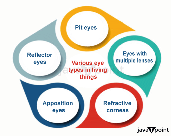
The six primary varieties of eyes found in living beings are as follows. These are divided into two categories: compound eyes and simple eyes. Simple eyes feature just one kind of connected ocular structure. Compound eyes may have numerous eye structures or be comprised entirely of photoreceptors. Human eyes may be categorized into several different eye kinds. The leading eye kinds found in living things are pit eyes, reflector eyes, apposition eyes, eyes with refractive corneas, eyes with multiple lenses, and superposition eyes. The many eye kinds found in living beings are listed below.
Types of Eye Shapes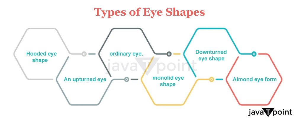
Next TopicStress Definition in Psychology
|
 For Videos Join Our Youtube Channel: Join Now
Feedback
- Send your Feedback to [email protected]
Help Others, Please Share


